
Medical image processing apparatus, medical image processing method, and medical image processing program

a technology of image processing and image processing program, which is applied in the field of medical image processing apparatus and medical image processing method, can solve the problems of inability to correct density unevenness in images, and may occur not only in mri images but also in ct images, so as to accurately compare target parts

Active Publication Date: 2021-11-02
FUJIFILM CORP
View PDF18 Cites 0 Cited by
  • Summary
  • Abstract
  • Description
  • Claims
  • Application Information

AI Technical Summary

Benefits of technology

[0011]The present invention has been made in view of the above circumstances, and it is an object of the present invention to make it possible to match the overall densities of two medical images and to correct density unevenness.
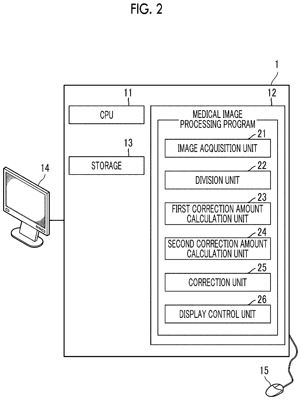
[0025]According to the present invention, the target parts included in the first medical image and the second medical image are divided into a plurality of regions corresponding to each other, and the correction amount for matching the density characteristics of each of the plurality of regions in the first medical image with density characteristics of corresponding regions in the second medical image is calculated as the first correction amount between the pixel value of the first reference pixel included in each of the plurality of regions in the first medical image and the pixel value of the second reference pixel corresponding to the first reference pixel for each of the plurality of regions in the second medical image. Then, the second correction amount for matching the first other pixel values other than the first reference pixel included in each of the plurality of regions in the first medical image with the pixel values of the second other pixels corresponding to the first other pixels for each of the plurality of regions in the second medical image is calculated based on the first correction amount. In addition, at least one of the first medical image or the second medical image is corrected based on the first correction amount and the second correction amount. For this reason, even in a case where the first medical image and the second medical image include unevenness of different pixel values, that is, density unevenness, not only the overall densities of the first medical image and the second medical image but also the density unevenness of the first medical image and the density unevenness of the second medical image can be matched with each other. Therefore, by using the corrected first medical image and second medical image, it is possible to accurately compare the target parts included in the first medical image and the second medical image with each other.

Problems solved by technology

However, it is not possible to correct density unevenness occurring in the images.
In addition, density unevenness may occur not only in MRI images but also in CT images and the like.

Method used


Image

  • Medical image processing apparatus, medical image processing method, and medical image processing program
  • Medical image processing apparatus, medical image processing method, and medical image processing program
  • Medical image processing apparatus, medical image processing method, and medical image processing program

Examples


Embodiment Construction

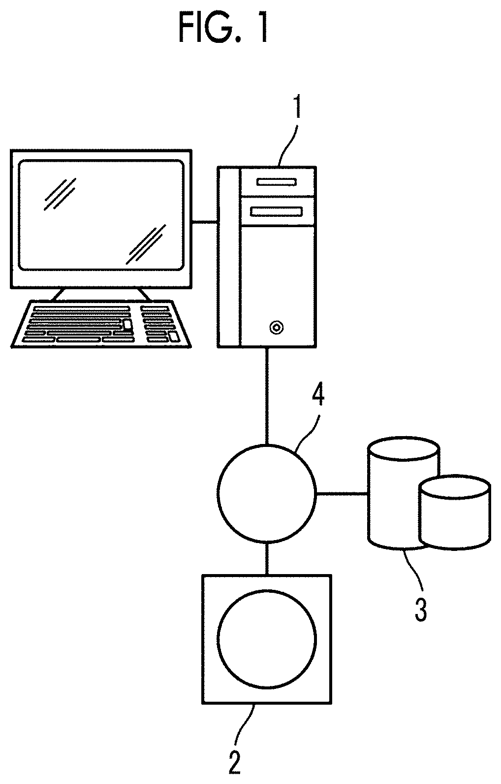
[0032]Hereinafter, embodiments of the present invention will be described with reference to the accompanying diagrams. FIG. 1 is a hardware configuration diagram showing the outline of a diagnostic support system to which a medical image processing apparatus according to an embodiment of the present invention is applied. As shown in FIG. 1, in the diagnostic support system, a medical image processing apparatus 1 according to the present embodiment, a three-dimensional image capturing apparatus 2, and an image storage server 3 are communicably connected to each other through a network 4.

[0033]The three-dimensional image capturing apparatus 2 is an apparatus that generates a three-dimensional image showing a part, which is a diagnostic target part of a patient who is a subject, as a medical image by imaging the part. Specifically, the three-dimensional image capturing apparatus 2 is a CT apparatus, an MRI apparatus, a PET apparatus, or the like. The medical image generated by the thre...


PUM


Abstract

A medical image processing apparatus, having a processor configured to divide brains included in a brain image and a standard brain image into a plurality of regions corresponding to each other, calculate a first correction amount between the pixel value of a first reference pixel included in each of the plurality of region in the brain image and the pixel value of a second reference pixel and a second correction amount for matching first other pixel values other than the first reference pixel included in each of the plurality of regions in the brain image with pixel values of second other pixels, and correct the brain image based on the first correction amount and the second correction amount.

Description

CROSS-REFERENCE TO RELATED APPLICATIONS[0001]This application is a Continuation of PCT International Application No. PCT / JP2018 / 020700 filed on May 30, 2018, which claims priority under 35 U.S.C. § 119(a) to Japanese Patent Application No. 2017-163010 filed on Aug. 28, 2017. Each of the above application(s) is hereby expressly incorporated by reference, in its entirety, into the present application.BACKGROUND OF THE INVENTION1. Field of the Invention[0002]The present invention relates to a medical image processing apparatus, a medical image processing method, and a non-transitory computer readable medium for storing a medical image processing program for correcting the density of a medical image.2. Description of the Related Art[0003]With the arrival of an aging society, the number of patients with dementia is increasing year by year. Dementia develops in a case where a protein called amyloid β accumulates in the brain and accordingly brain atrophy progresses and cognitive ability d...

Claims


Application Information

Patent Timeline
Patent Type & Authority Patents(United States)
IPC IPC(8): A61B5/00A61B5/055A61B6/03G06T5/00G06T5/40G06T5/50G06T7/11
CPCG06T5/40A61B5/055G06T5/50G06T7/11A61B2576/026G06T2207/10088G06T2207/30016A61B5/0042A61B6/03G06T5/008
Inventor WANG, CAIHUA
Owner FUJIFILM CORP
Learn More