
Method for determining the haemoglobin F content of an erythroid cell

a technology of erythroid cells and haemoglobin, which is applied in the direction of fluorescence/phosphorescence, instruments, material analysis, etc., can solve the problems of increasing the incidence of children's mortality in developing countries, increasing the mean hbf content, and not being sensitive to methods

Active Publication Date: 2022-01-25
UNIV PARIS EST CRETEIL VAL DE MARNE +3
View PDF4 Cites 0 Cited by
  • Summary
  • Abstract
  • Description
  • Claims
  • Application Information

AI Technical Summary

Benefits of technology

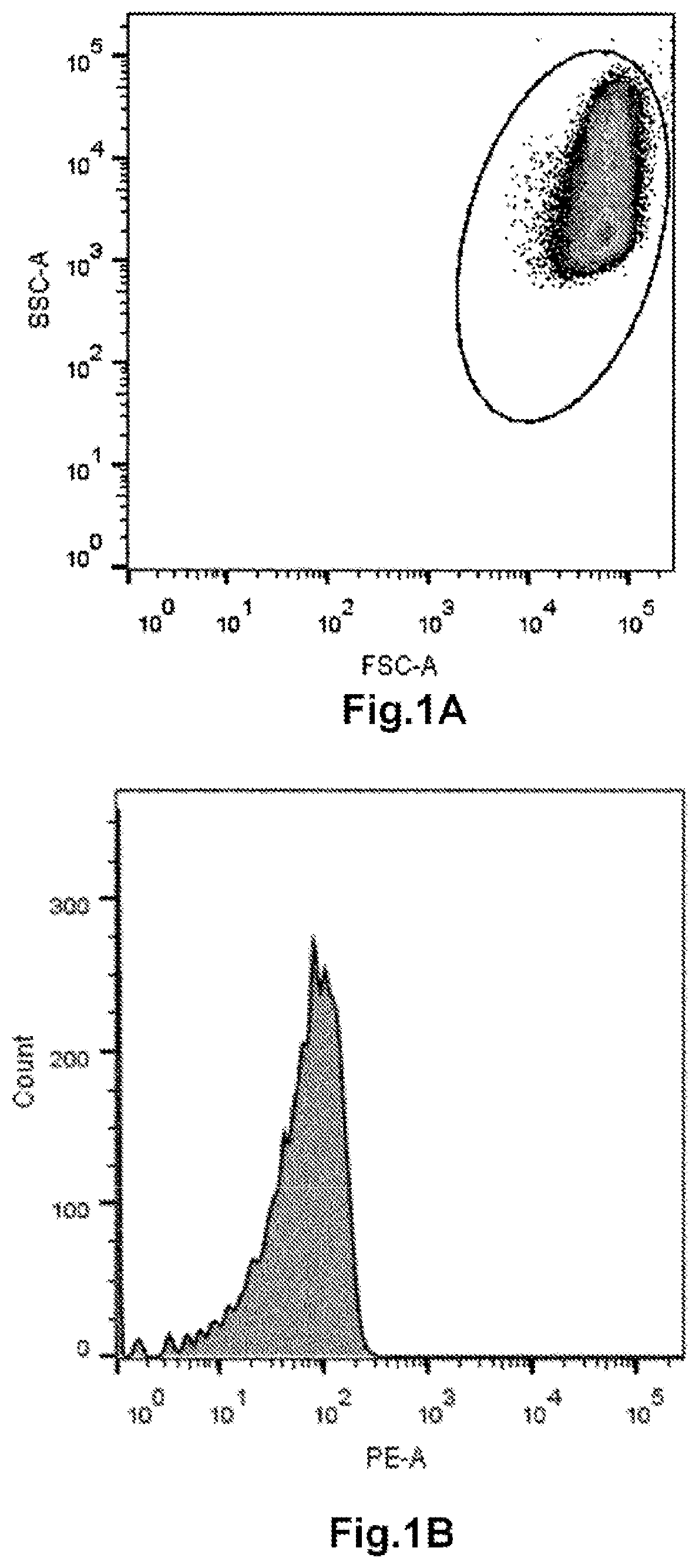
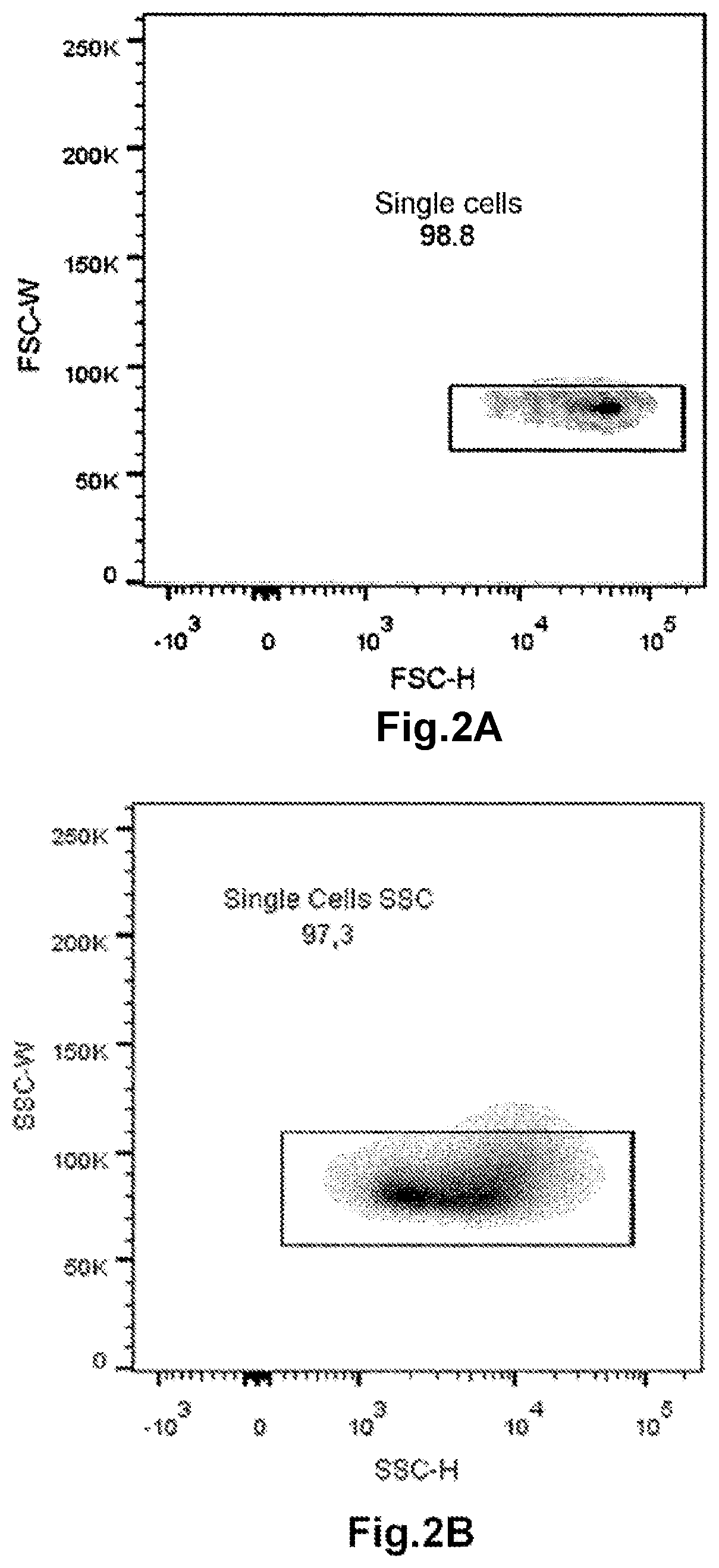
[0051]The inventors have demonstrated that flow cytometry is a particularly suitable technique for easily and accurately determining the HbF content of each erythroid cell of a set of erythroid cells.
[0060]Generally, in one particular embodiment, the membrane of the isolated erythroid cells is fixed before the permeabilization step. The fixing of the erythroid cell membrane makes it possible to prevent (or to limit) the lysis of the erythroid cells during the permeabilization step. Several chemical compounds are at the disposal of those skilled in the art for fixing the membrane of the erythroid cells. In one particular embodiment, the membrane of the isolated erythroid cells is fixed with sodium azide and / or formaldehyde. Generally, the sodium azide and / or formaldehyde are simply added to a pellet of erythroid cells or to a suspension of erythroid cells.
[0061]Step c) consists in labeling the HbF of the erythroid cells obtained in step b) (that is to say the isolated erythroid cells of which the membrane is permeabilized) with an anti-HbF antibody conjugated to a fluorochrome (also hereinafter referred to as “anti-HbF antibody”). Advantageously, step c) is carried out under conditions which allow intracellular binding between the anti-HbF antibody and the HbF. This step makes it possible to obtain “labeled erythroid cells”. Preferably, a sufficient amount of anti-HbF antibody to be able to label all of the HbF is used, for example an excess of anti-HbF antibody is used. In particular, the step c) consists in labeling the HbF of the erythroid cells obtained in step b) with an anti-HbF antibody conjugated to a fluorochrome under conditions which allow intracellular binding between the anti-HbF antibody and the HbF. That is to say that the binding between the anti-HbF antibody and the HbF takes place inside the erythroid cell. Generally, the labeling consists in incubating the erythroid cells, the membrane of which was permeabilized, with the anti-HbF antibody for a period of time sufficient to allow intracellular binding between the anti-HF antibody and the HbF. The period of time is generally a few minutes, for example between 5 min and 20 min.
[0062]In one particular embodiment, the erythroid cells are washed after the labeling step. The washing makes it possible to eliminate the anti-HbF antibodies not bound to the HbF. The washing can be carried out with a suitable buffer, for example a PBS buffer. The washing also makes it possible to eliminate the chemical agent that was used to permeabilize the membrane of the red blood cells. The red blood cells thus washed are then used in the flow cytometry measuring step.
[0083]Advantageously, the amount of transfused red blood cells makes it possible to determine a percentage of transfused red blood cells relative to the total red blood cells of the patient. This makes it possible in particular to monitor the change in the amount of transfused red blood cells (or in the percentage of transfused red blood cells) and thus to assist in the diagnosis in certain particular cases, for example the distinction between allo antibodies and auto antibodies directed against red blood cells, which may be transfused or the patient's red blood cells.Monitoring of the Therapeutic Efficacy for Sickle Cell Disease or for β-Thalassemia
[0088]Thus, it is possible to monitor the progression in the therapeutic efficacy of a treatment, aimed at increasing HbF, for sickle cell disease or for β-thalassemia over time, before, during or after the treatment of the patient. It is thus possible to monitor the progression of the therapeutic efficacy of known treatments, or of new treatments. It is thus possible to identify new treatments for sickle cell disease or for β-thalassemia which make it possible to increase HbF synthesis in red blood cells.

Problems solved by technology

However, the lack of early screening and the limited access to quality treatments and care continue to cause the incidences of mortality in children to increase in developing countries.
Nevertheless, the increase in the mean HbF content is not always associated with a clinical improvement in sickle cell disease patients.
Although widely used in the diagnosis of fetal-maternal incompatibilities and in the understanding of hemoglobinopathies, these methods are not very sensitive and are limited to a qualitative measurement of F cells (red blood cells positive by immunofluorescence microscopy, immunodiffusion or by flow cytometry, regardless of the HbF expression level).

Method used


Image

  • Method for determining the haemoglobin F content of an erythroid cell
  • Method for determining the haemoglobin F content of an erythroid cell
  • Method for determining the haemoglobin F content of an erythroid cell

Examples


example 1

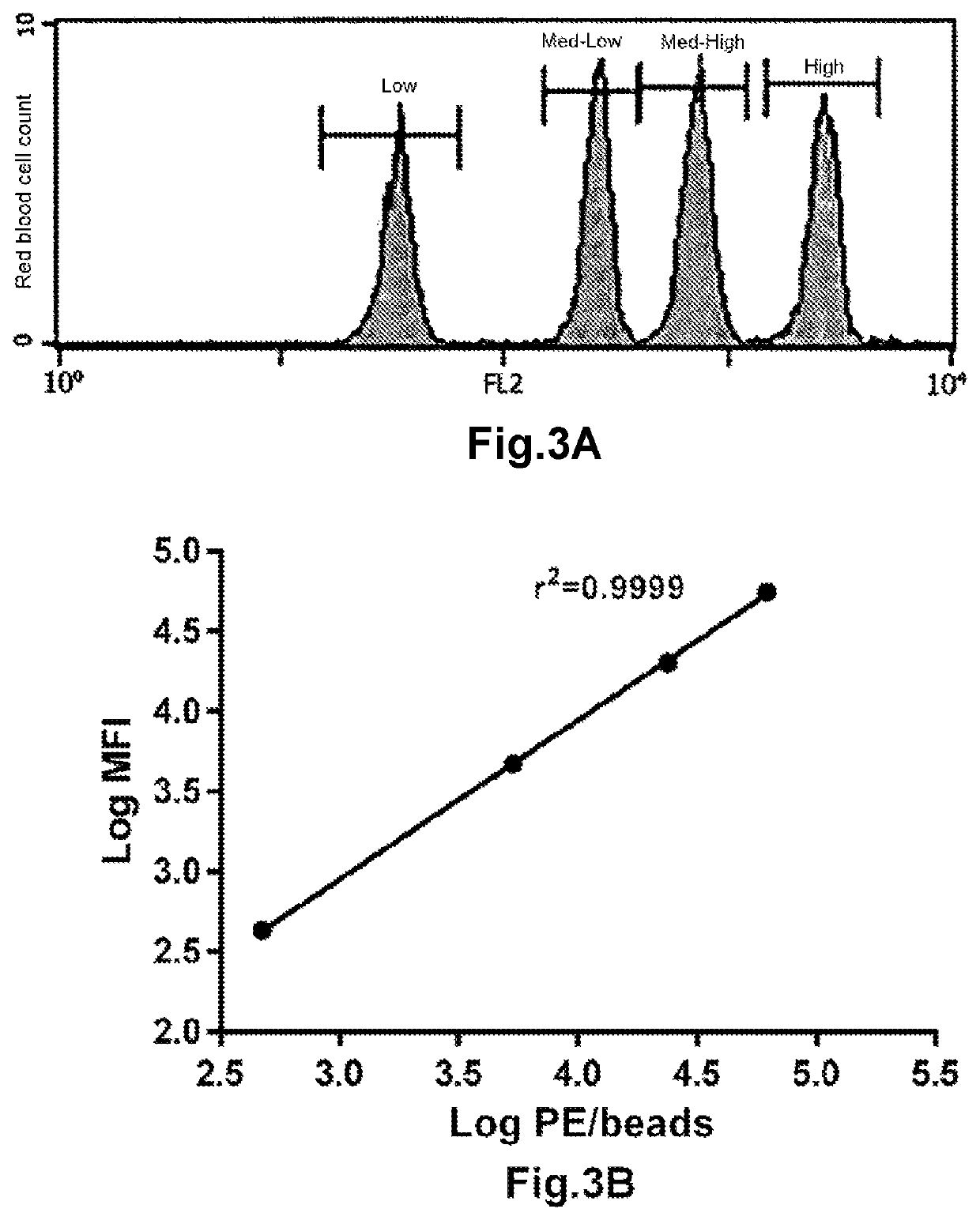
nt of a Method for Determining the HbF Content of Each Red Blood Cell: Construction of a Standard Curve

[0113]A group of patients having a perfectly homogeneous distribution of the HbF content of each red blood cell was selected from the SICLOPEDIE collection monitored at the Unite de Maladies Génétiques du Globule Rouge [Red Blood Cell Genetic Diseases Unit] at the Centre Hospitalier Universitaire Henri Mondor [Henri Mondor University Hospital Center] in Creteil. This group is composed of adult patients (age≥18 years old) having a hereditary persistence of HbF (HPFH) or intermediate or minor β-thalassemia, and an HbF content of each of their red blood cells which is homogeneous and constant over time. The HbF content of the total red blood cells of patients was determined, in the context of patient monitoring, by regular measurement of the % HbF (by HPLC) and of the mean HbF content per red blood cell (MCHbFCo). Patients having a major sickle cell syndrome (SS, Sβ-Thalassemia or SC)...

example 2

n of the Method for Determining the HbF Content of Each Red Blood Cell

1. Characteristics of the Included Patients

[0157]A cohort of 12 patients was selected in order to constitute a range of HbF content, the criterion for selecting these patients was the confirmation of a homogeneous distribution, Log-Normal of their HbF. The data from these patients were used to construct a standard curve (cf. example 1, point 5) serving to determine the HbF content of each red blood cell. Table 2 summarizes the demographic data and the genetic characteristics of these patients.

[0158]

TABLE 2Demographic and biological data of the includedpatients for constructing the standard curveAge atinclusionGeneticHbFMCHCoMCHbFCoMCVPatients(years)Sexcharacteristics(%)(pg)(pg)(μm3)118Maleβ-thalassemia10024.524.573262Maleβ-thalassemia69.627.218.9391350Maleβ-thalassemia520.112.3664446Maleβ-thalassemia53.219.310.2763550FemaleGhanaian HPFH234.80289.7481639Femaleβ-thalassemia27.9287.7681739Maleβ-thalassemia2123.857284...

example 3

the HbF Distribution During the Treatment with Hydroxyurea

1. Patients

[0167]A monocentric, longitudinal, prospective study was carried out on a cohort of 29 adult sickle cell disease patients (age≥18 years old) exhibiting an SS homozygotes mutation, beginning a treatment with hydroxyurea, regularly monitored at the major sickle cell syndrome reference Center at the Centre Hospitalier Universitaire Henri Mondor [Henri Mondor University Hospital Center] in Creteil. Patients having had an attack requiring hospitalization and / or a transfusion exchange program during the previous 3 months before inclusion and also patients who were pregnant or who were wanting to get pregnant were excluded from the study.

[0168]The patients exhibiting the eligibility criteria were monitored at various times before and during the treatment with hydroxyurea: D0 (before the beginning of the treatment with hydroxyurea), at 15 days, 1 month, 3 months, 4 months and 6 months or more after the setting up of the tr...


PUM

PropertyMeasurementUnit
timeaaaaaaaaaa
timeaaaaaaaaaa
volumeaaaaaaaaaa

Abstract

The invention concerns a method for determining, by flow cytometry, the hemoglobin F (HbF) content of each erythroid cell of a set of erythroid cells. This method applies in particular to determining the HbF content of each red blood cell of a set of red blood cells. The invention also concerns a method for determining the amount of red blood cells transfused into a patient and for monitoring the therapeutic efficacy of a treatment for sickle cell disease or β-thalassemia.

Description

CROSS-REFERENCE TO RELATED APPLICATIONS[0001]The present application is a U.S. National Stage Entry of International Application No. PCT / FR2017 / 053015, filed Nov. 3, 2017, which claims priority to French Patent Application No. 1660713, filed Nov. 4, 2016.TECHNICAL FIELD[0002]The invention relates to a novel method for determining, in vitro, the hemoglobin F (HbF) content of each erythroid cell of a set of erythroid cells, preferably of each red blood cell of a set of red blood cells, by flow cytometry. The invention also relates to a novel method for determining, in vitro, the amount of red blood cells transfused into a patient and to a method for monitoring the therapeutic efficacy of a treatment for sickle cell disease or for β-thalassemia.TECHNOLOGICAL BACKGROUND[0003]Sickle cell disease is a genetic disease of hemoglobin which was historically characterized by the deformation of red blood cells (RBCs) in response to a decrease in oxygen partial pressure. The red blood cells lose...

Claims


Application Information

Patent Timeline
Patent Type & Authority Patents(United States)
IPC IPC(8): G01N33/72G01N33/80G01N33/49G01N21/64G01N15/14G01N15/00G01N15/10
CPCG01N33/721G01N15/1459G01N21/6428G01N21/6486G01N33/80G01N33/49G01N2015/0073G01N2015/1006G01N2800/52
Inventor CAMBOT, MARIEVANDEMEULEBROUCK, GAETANANOIZAT PIRENNE, FRANCEBARTOLUCCI, PABLORAKOTOSON, MARIE GEORGINEGALACTEROS, FRÉDÉRICHEBERT, NICOLAS
Owner UNIV PARIS EST CRETEIL VAL DE MARNE
Learn More