
Attenuation of fibroblast proliferation
- Summary
- Abstract
- Description
- Claims
- Application Information
AI Technical Summary
Benefits of technology
Problems solved by technology
Method used
Image

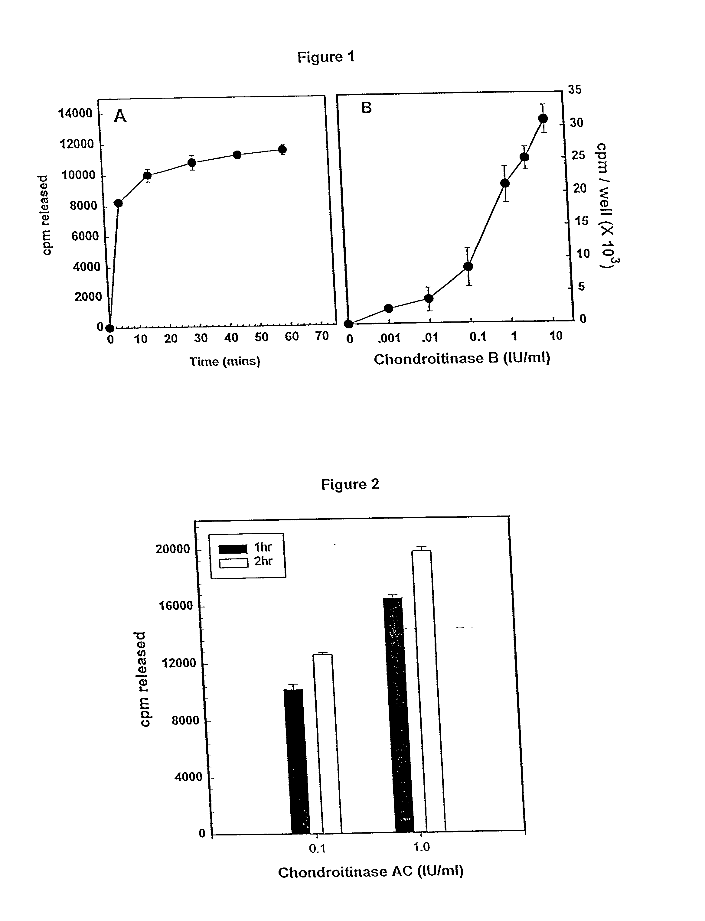
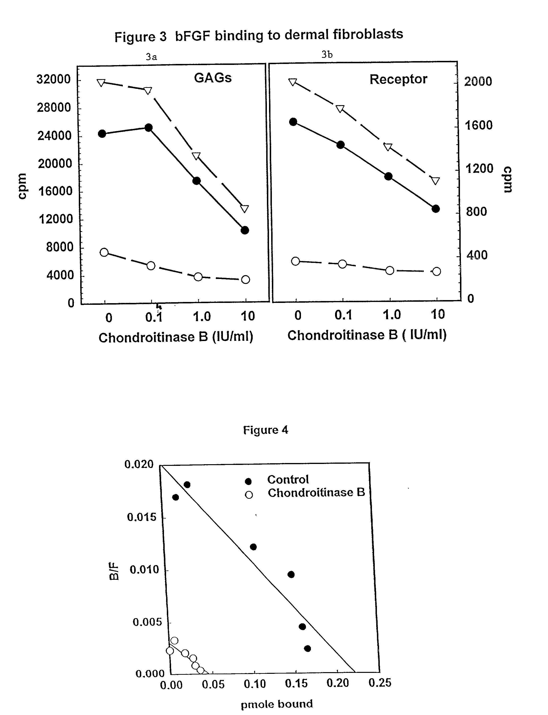
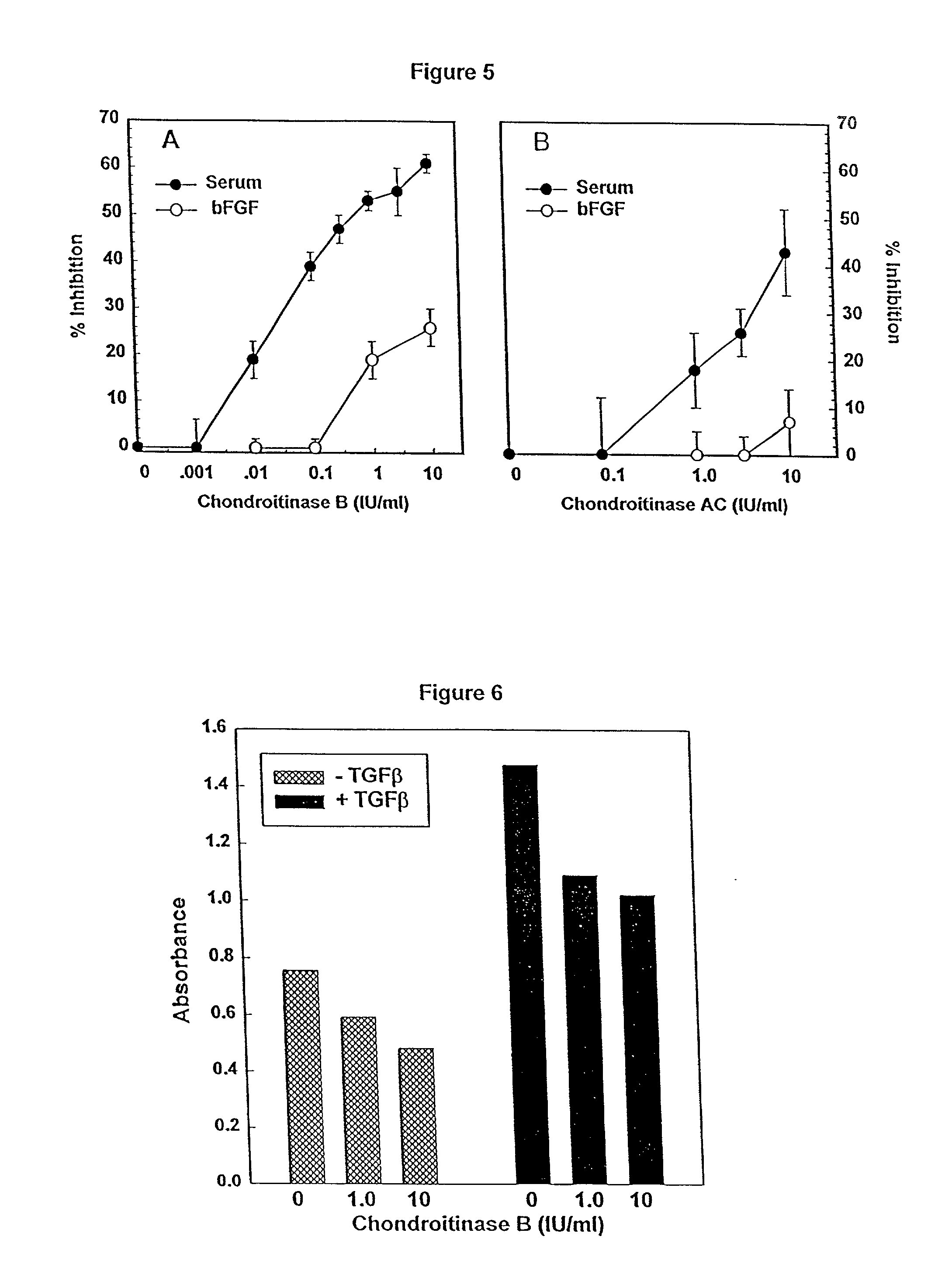
Examples
example 1
[0048] Chondroitinase B (no EC number) and chondroitinase AC (EC 4.2.2.5) are recombinant proteins expressed in Flavobacterium heparinum (PCT / US95 / 08560 "Chondroitin Lyase Enzymes"). Specific activity and substrate specificity were determined for each enzyme, using a kinetic spectrophotometric assay, performed essentially as described in PCT / US95 / 0856. In these assays, enzyme concentrations were 0.25 IU / ml and substrate concentrations were 0.5 mg / ml (chondroitin sulfate B and chondroitin sulfate AC) or 0.75 mg / ml (heparan sulfate). The specific activities of the enzymes were: 97 IU / mg for Chondroitinase B and 221 IU / mg for chondroitinase AC.
[0049] The substrate specificity of ultra-purified Chondroitinase B and AC were determined by testing the ability of the enzymes to degrade chondroitin sulfate B, chondroitin sulfate A, chondroitin sulfate C, and heparan sulfate. As shown in Table 1, both enzymes were active towards the corresponding sulfated glycosami...
example 2
Removal of Glycosaminoglycans from Cells
[0051] The effectiveness of the chondroitinases B and AC in removing sulfated glycosaminoglycans from cells was examined using cells with glycosaminoglycans labeled by incubation with .sup.35S. Fibroblasts were plated at a density of 6.times.10.sup.4 cells / well in 24 well plates, in DMEM with 10% serum. After 24 hrs, medium was changed to Fisher's medium containing 10% serum and 25 microCi / ml of Na.sub.2.sup.35SO.sub.4, and incubation continued for 2.5 days. The medium was removed and cells rinsed 2.times. with DMEM then treated with Chondroitinase B or AC as indicated. Medium was removed and radioactivity determined. The release of sulfated glycosaminoglycans from cells by enzyme was expressed as cpm / well of chondroitinase-treated cells minus cpm / well of untreated cells.
[0052] Cells were exposed to 1.0 IU / ml of Chondroitinase B, at 37.degree. C. for variable lengths of time. As shown in FIG. 1A, maximal release of sulfated GAGs by chondroitin...
example 3
Effects on bFGF Binding
[0054] Iodinated bFGF was obtained from Dupont NEN, specific activity greater than 1200 Ci / mmol. Fibroblasts were plated in 48 well dishes and grown to confluence. Prior to binding assays, cells were treated with medium or enzyme as indicated for proliferation assays. Following enzyme treatments, cells were chilled and binding assays carried out at 4.degree. C. Cells were incubated for one hour with 25 ng / ml of .sup.125l-bFGF alone, or with the addition of 25 microg / ml of unlabelled bFGF in binding buffer (DMEM, 25 mM HEPES, 0.05% gelatin). Following incubation with bFGF, cells were washed 2.times. with ice cold binding buffer. Glycosaminoglycan-bound .sup.125l-bFGF was removed with two rinses with wash buffer (2M NaCl in 20 mM HEPES, pH 7.4). Receptor bound .sup.125I-bFGF was removed by washing 2.times. with wash buffer (pH 4.0) (Fannon and Nugent (1996) J. Biol. Chem. 271:17949-17956).
[0055] Numerous growth factors have been shown to bind to heparan sulfate ...
PUM
| Property | Measurement | Unit |
|---|---|---|
| Disorder | aaaaa | aaaaa |
Abstract
Description
Claims
Application Information