Spinal ligament modification

Active Publication Date: 2006-02-16
VERTOS MEDICAL
View PDF97 Cites 125 Cited by
  • Summary
  • Abstract
  • Description
  • Claims
  • Application Information

AI Technical Summary

Benefits of technology

[0025] In yet further other embodiments, a method for preventing leakage of cerebrospinal fluid from an opening in a thecal sac in a spine may comprise accessing the epidural space in the vicinity of the opening and inserting a volume of fluid into the epidural space, where the fluid thickens as it attains body temperature such that the fluid blocks the opening in the thecal sac.
[0026] In further embodiments, a bone cutting device can be used to access the ligamentum flavum and epidural space, to perform a laminotomy or to allow placement of a cannula. Using a cannula fixed within (exten

Problems solved by technology

The excessive epidural fat compresses the dural sac, nerve roots and blood vessels contained therein and resulting in back and leg pain and weakness and numbness of the legs.
These conservative treatment options frequently fail.
Also, because these spine stabilizing back muscles and ligaments are stripped and cut off, the spine these patients frequently develop spinal instability post-operatively.
Percutaneous discectomy devices with fluoroscopic guidance have been used successfully to treat disorders of the disc but not to treat spinal stenosis or the liga

Method used


Image

  • Spinal ligament modification
  • Spinal ligament modification
  • Spinal ligament modification

Examples


Embodiment Construction

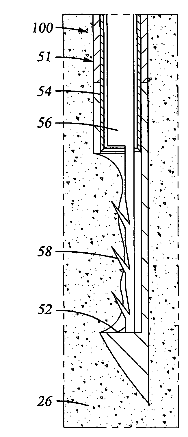
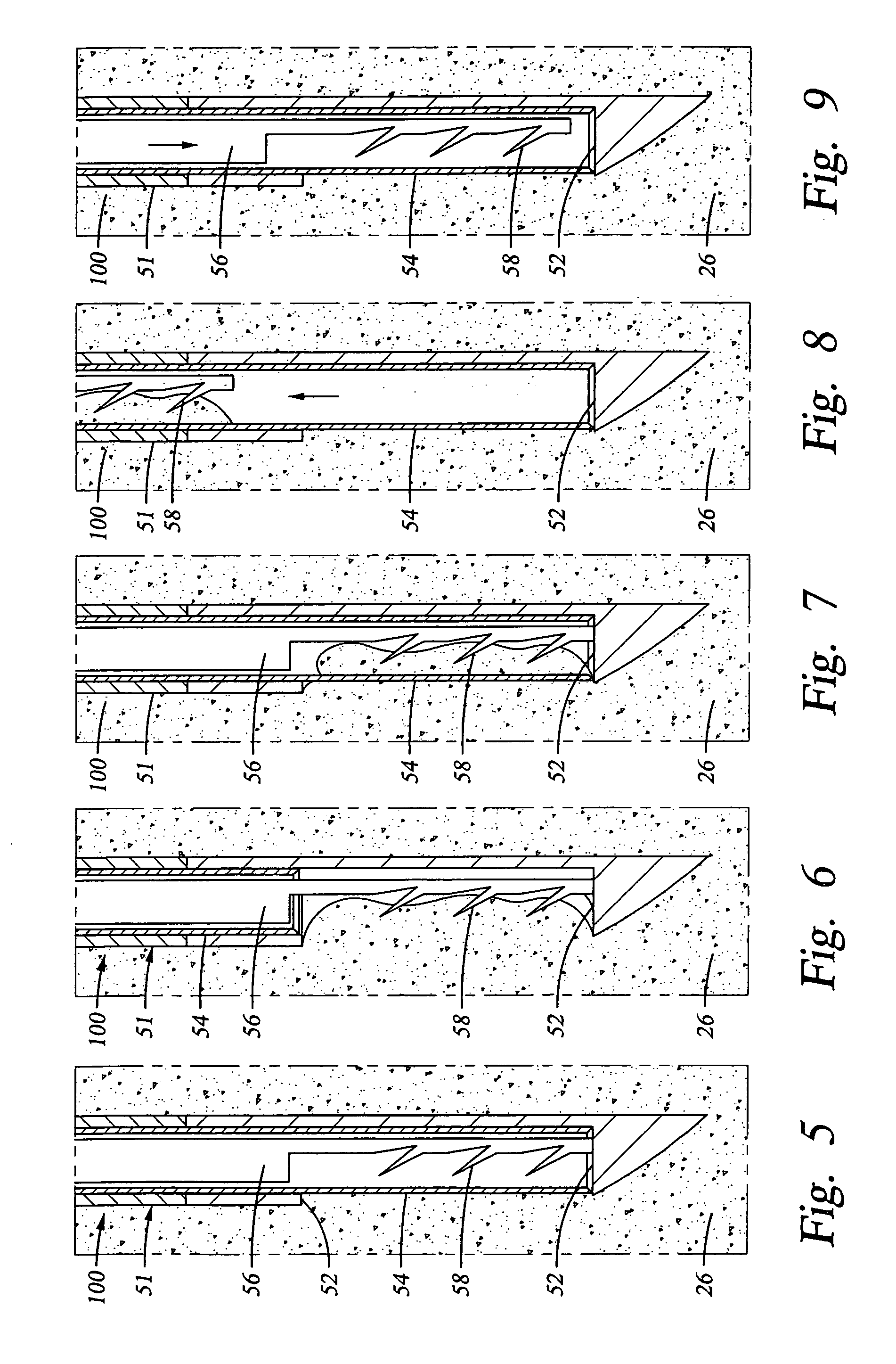
[0046] The epidural space is the space between the ligamentum flavum and the thecal sac. This space is filled with blood vessels and fat. The nerves contained within the thecal sac are normally surrounded by cerebrospinal fluid (CSF). When the ligamentum flavum hypertrophies, the blood vessels that supply the nerves of the cauda equina are compressed. This results in ischemic pain termed spinal claudication. The nerve roots may also be compressed resulting in back and / or leg pain.

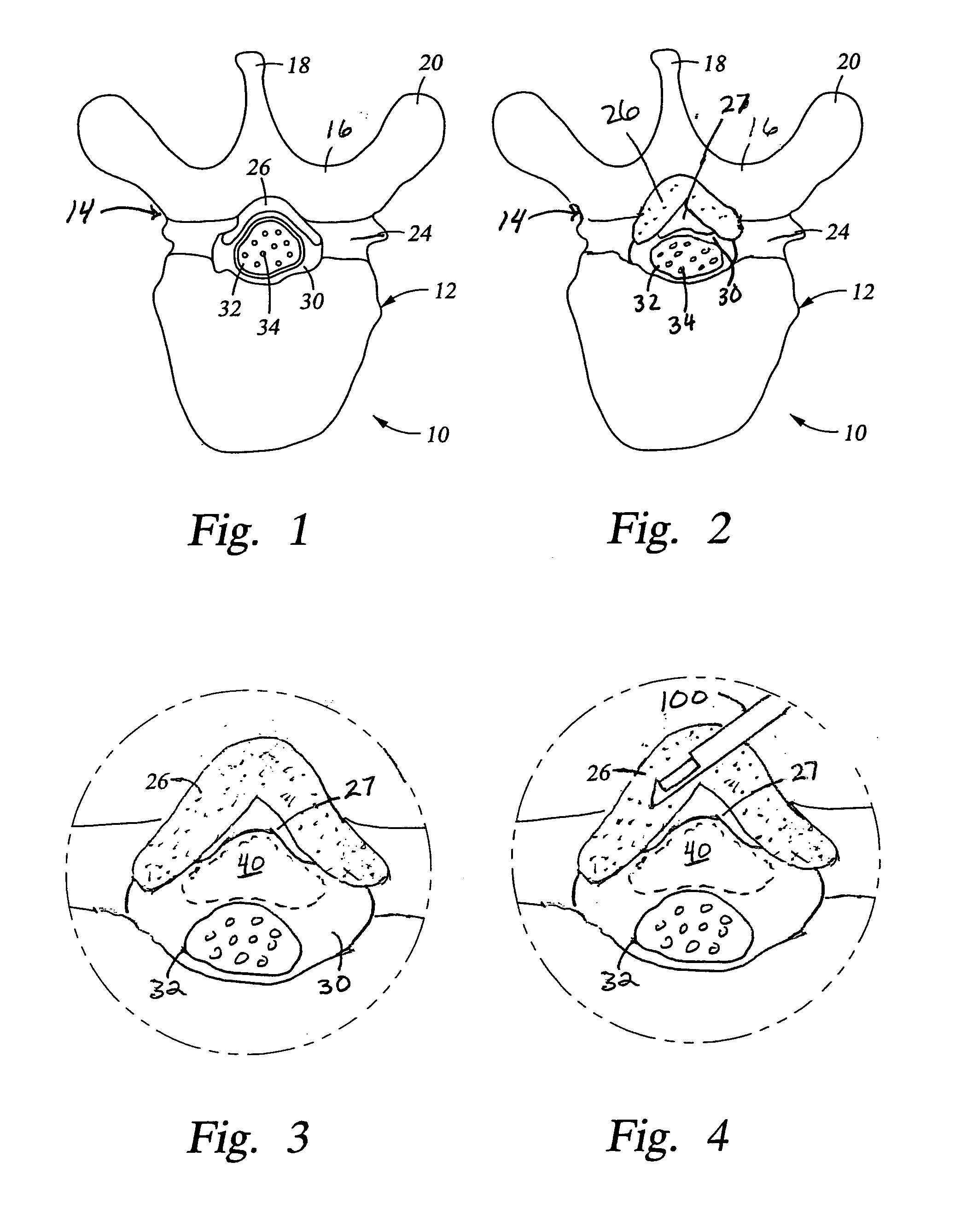
[0047] Referring again to FIG. 1, the posterior border of the normal epidural space 30 is formed by the normally thin ligamentum flavum 26 and posterior epidural fat (not shown). Ligamentum flavum 26 extends from the lamina above the interspinous space to the lamina below the interspinous space. The dural sleeve (thecal sac) 32 contains nerve roots 34 surrounded by cerebrospinal fluid. The nerve roots 34 normally comprise only a small proportion of the thecal sac volume.

[0048] In FIG. 2, spinal stenosis i...


PUM


Abstract

A method for treating stenosis in a spine comprises percutaneously accessing the epidural space in a stenotic region of interest, compressing the thecal sac in the region of interest to form a safety zonem, inserting a tissue removal tool into tissue in the working zone, using the tool to percutaneously reduce the stenosis; and utilizing imaging to visualize the position of the tool during at least a part of the reduction step. A tissue excision system for performing percutaneous surgery, comprises a cannula comprising a tissue-penetrating member having a distal end defining an aperture on one side thereof, an occluding member slidably received on or in the cannula and closing the aperture when the occluding member is adjacent the cannula distal end, means for engaging adjacent tissue via the aperture, and cutting means for resecting a section of the engaged tissue.

Description

CROSS-REFERENCE TO RELATED APPLICATIONS [0001] This application claims benefit of U.S. application Ser. No. 60 / 592,099 filed Jul. 29, 2004, entitled “Device for Percutaneous Treatment of Spinal Stenosis,” which is incorporated herein by reference in its entirety.TECHNICAL FIELD OF THE INVENTION [0002] The present invention relates to a minimally invasive method, device and system for treating spinal disorders using imaging guidance. This invention also relates to devices used to reduce stenosis and increase the cross-sectional area of the spinal canal and to devices used to treat excess fat within the spinal canal or epidural lipomatosis. This invention also relates to methods, devices, therapies and medications used to treat disorders that involve the epidural space. BACKGROUND OF THE INVENTION [0003] The spine comprises a stack of vertebrae with an intervertebral disc between adjacent vertebrae. As shown in FIG. 1, each vertebra 10 includes a vertebral body 12 that supports a bony...

Claims


Application Information

Patent Timeline
IPC IPC(8): A61B17/32
CPCA61B10/0233A61B2017/320064A61B10/0283A61B17/0401A61B17/0482A61B17/1671A61B17/320016A61B17/32002A61B17/32053A61B17/320783A61B17/3401A61B17/3478A61B2010/045A61B2017/00261A61B2017/00349A61B2017/0412A61B2017/0419A61B2017/0427A61B2017/0437A61B2017/06052A61B2017/06176A61B2017/320008A61B2017/32004A61B10/0275
Inventor SOLSBERG, MURRAY D.SCHOMER, DONALD
Owner VERTOS MEDICAL
Why Eureka
Try Eureka