Surgical microscope having an OCT-system and a surgical microscope illuminating module having an OCT-system

Active Publication Date: 2008-05-22
CARL ZEISS MEDITEC AG
View PDF14 Cites 21 Cited by
  • Summary
  • Abstract
  • Description
  • Claims
  • Application Information

AI Technical Summary

Benefits of technology

[0022]It is possible to shift the OCT-scanning plane of the OCT-system relative to a viewing plane for optical viewing beam paths in that an adjustable collimating optic is provided in the

Problems solved by technology

An interference signal results

Method used


Image

  • Surgical microscope having an OCT-system and a surgical microscope illuminating module having an OCT-system
  • Surgical microscope having an OCT-system and a surgical microscope illuminating module having an OCT-system
  • Surgical microscope having an OCT-system and a surgical microscope illuminating module having an OCT-system

Examples


Embodiment Construction

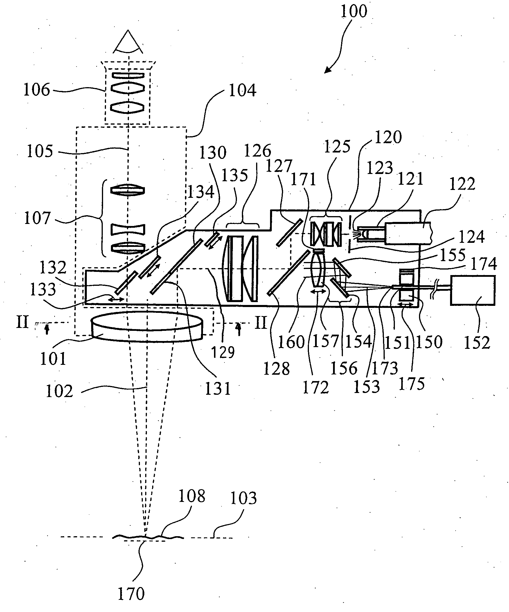
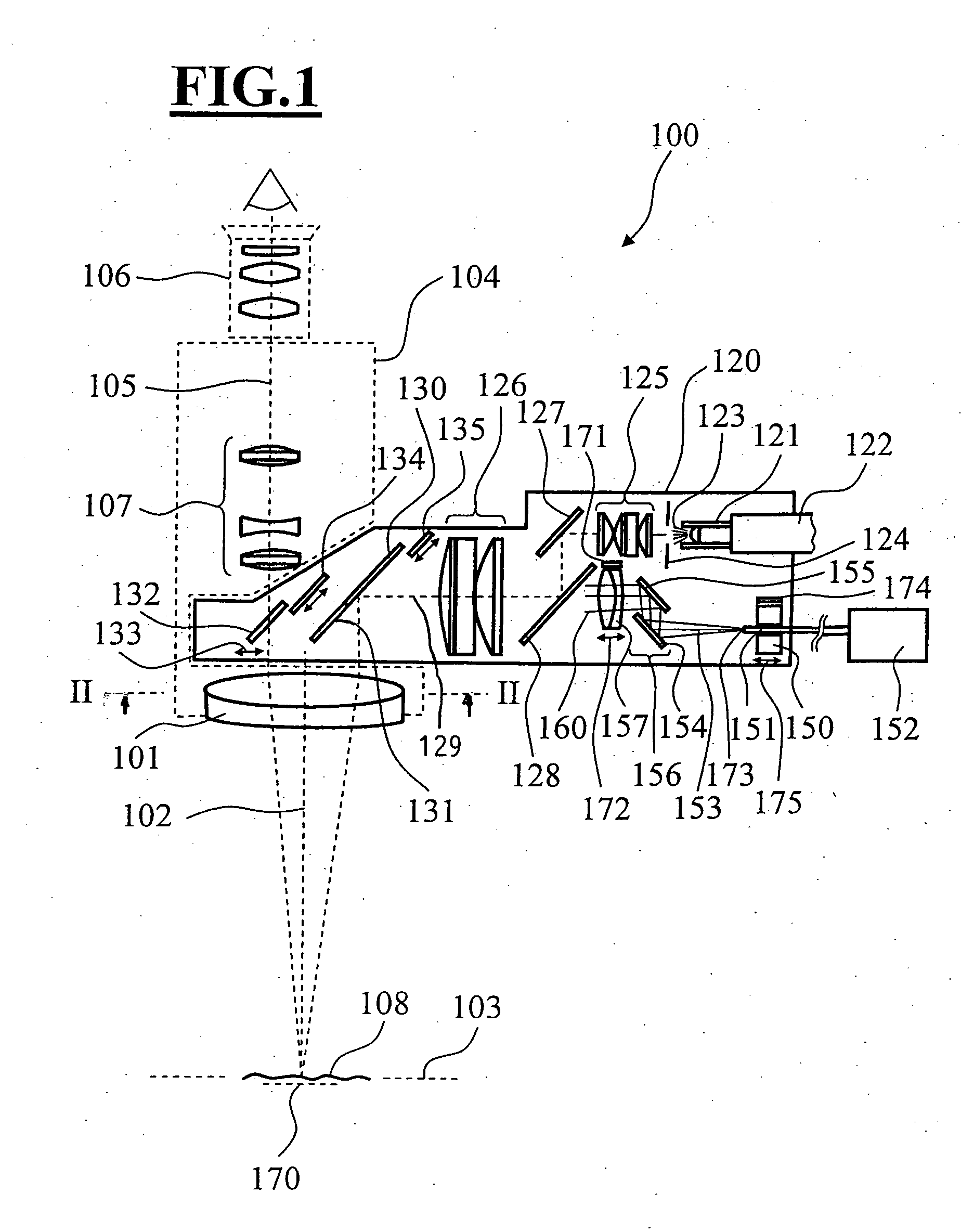
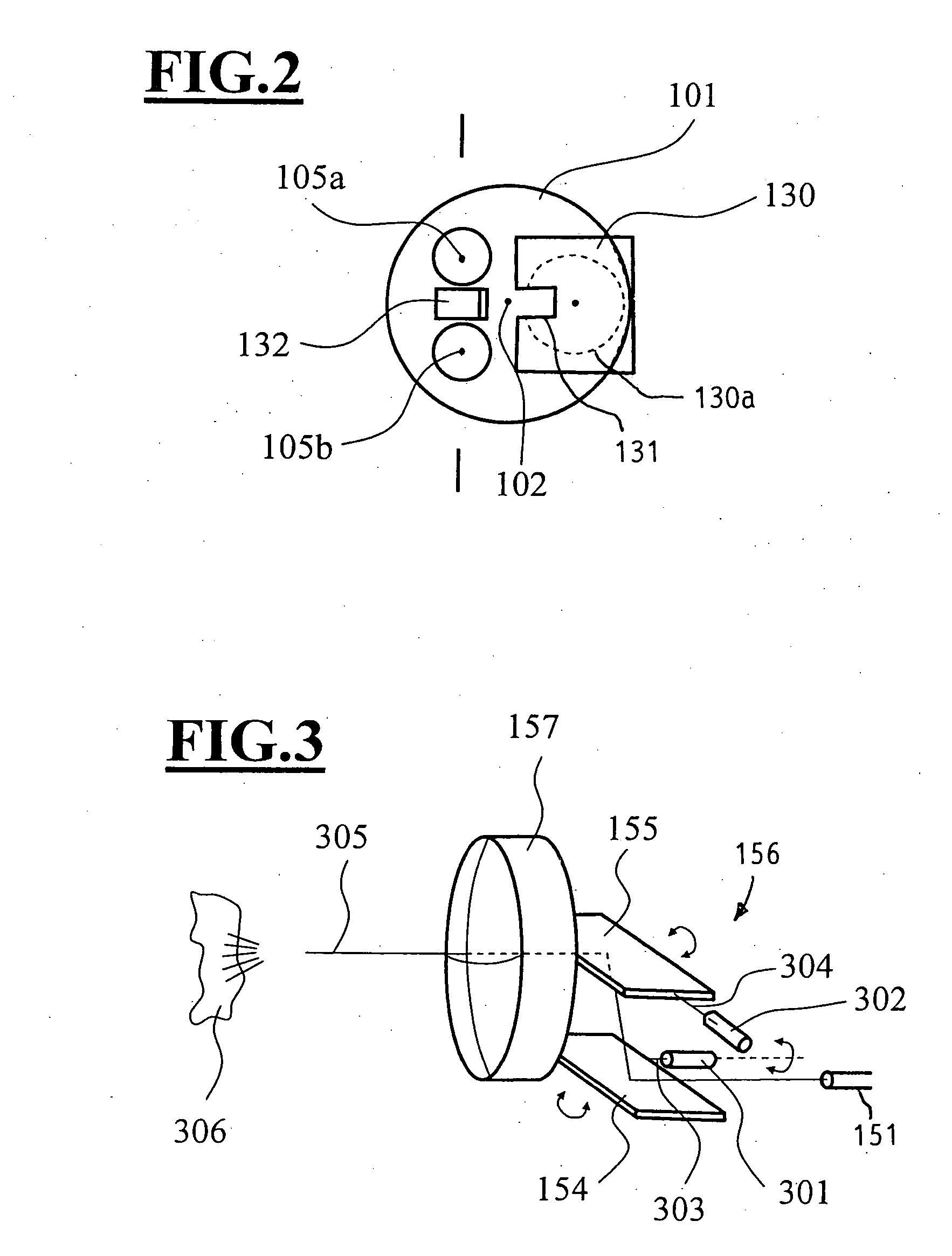
[0029]The surgical microscope 100 in FIG. 1 has a microscope main objective 101 defining an optical axis 102 as well as a focus plane 103. The microscope main objective 101 is accommodated in a surgical microscope base body 104. Stereoscopic viewing beam paths of a binocular tube 106 are represented by broken line 105 in FIG. 1 and pass through the microscope main objective 101. The surgical microscope 100 includes a zoomable magnification system 107.

[0030]The surgical microscope 100 has an illuminating unit in the form of an illuminating module 120 for illuminating the object region 108. This illuminating module 120 includes a receptacle 121 for a first light conductor 122 which provides illuminating light 123 from a light source (not shown). An adjustable field diaphragm 124 is illuminated with an illuminating light 123 exiting from the first light conductor 122. An illuminating optic is mounted in the illuminating module 120. The illuminating optic includes the following: a first...


PUM


Abstract

A surgical microscope (100) has an illuminating module (120). The illuminating module contains an illuminating optic which images a field diaphragm (124) to a parallel illuminating beam path at infinity. The field diaphragm (124) is illuminated by a light source. The illuminating optic includes a first lens assembly and a second lens assembly which functions to image the field diaphragm (124) into the object region (108) via the microscope main objective (101) of the surgical microscope (100). An in-coupling element (128) is provided between the first lens assembly (125) and the second lens assembly (126) and this in-coupling element couples the OCT-scanning beam into the illuminating beam.

Description

CROSS REFERENCE TO RELATED APPLICATIONS[0001]This application claims priority of German patent application nos. 10 2006 052 513.2 and 10 2007 019 677.8, filed Nov. 6, 2006 and Apr. 24, 2007, respectively, the entire contents of which are incorporated herein by reference.FIELD OF THE INVENTION[0002]The invention relates to a surgical microscope having a viewing beam path for examining a region of an object. The surgical microscope includes an illuminating system having an illuminating optic which images a field diaphragm illuminated by a light source to a parallel illuminating beam at infinity. The illuminating optic includes a first lens assembly and a second lens assembly and the surgical microscope includes an objective which is mounted in the parallel illuminating beam path in order to image the field diaphragm onto the object region. The surgical microscope also includes an OCT-system for examining the object region and the OCT-system includes an OCT-scanning beam which is condu...

Claims


Application Information

Patent Timeline
IPC IPC(8): G02B21/06
CPCA61B5/0066A61B5/0073A61B19/52G02B21/0032A61B19/5223G02B21/0012A61B19/5202A61B90/20A61B90/30A61B90/36
Inventor REIMER, PETERHAUGER, CHRISTOPHABELE, ALFONSSEESSELBERG, MARKUS
Owner CARL ZEISS MEDITEC AG
Why Eureka
Try Eureka