Method for classifying breast tissue density

a breast tissue density and density technology, applied in mammography, image analysis, image enhancement, etc., can solve the problems of increased difficulty in detection of cancer sites of denser breasts, less success of mammography imaging techniques with denser breast tissue than with predominantly fat tissue, etc., to achieve the effect of assisting mammogram image rendering and diagnosis

Inactive Publication Date: 2008-07-03
CARESTREAM HEALTH INC
View PDF7 Cites 32 Cited by
  • Summary
  • Abstract
  • Description
  • Claims
  • Application Information

AI Technical Summary

Benefits of technology

[0009]One object of the present invention is to provide an automated method for class

Problems solved by technology

For example, there is a consideration that mammographic imaging techniques are less successful with denser breast tissue than with predominantly fat tissue.
Fibro-glandula

Method used


Image

  • Method for classifying breast tissue density
  • Method for classifying breast tissue density
  • Method for classifying breast tissue density

Examples


Embodiment Construction

[0023]The following is a detailed description of the preferred embodiments of the invention, reference being made to the drawings in which the same reference numerals identify the same elements of structure in each of the several figures.

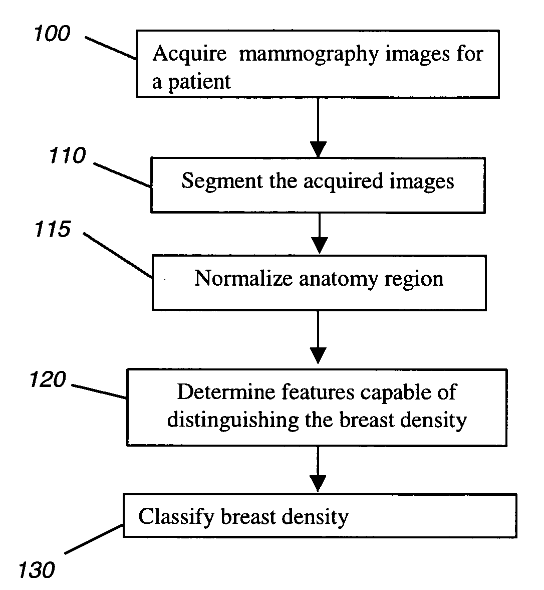
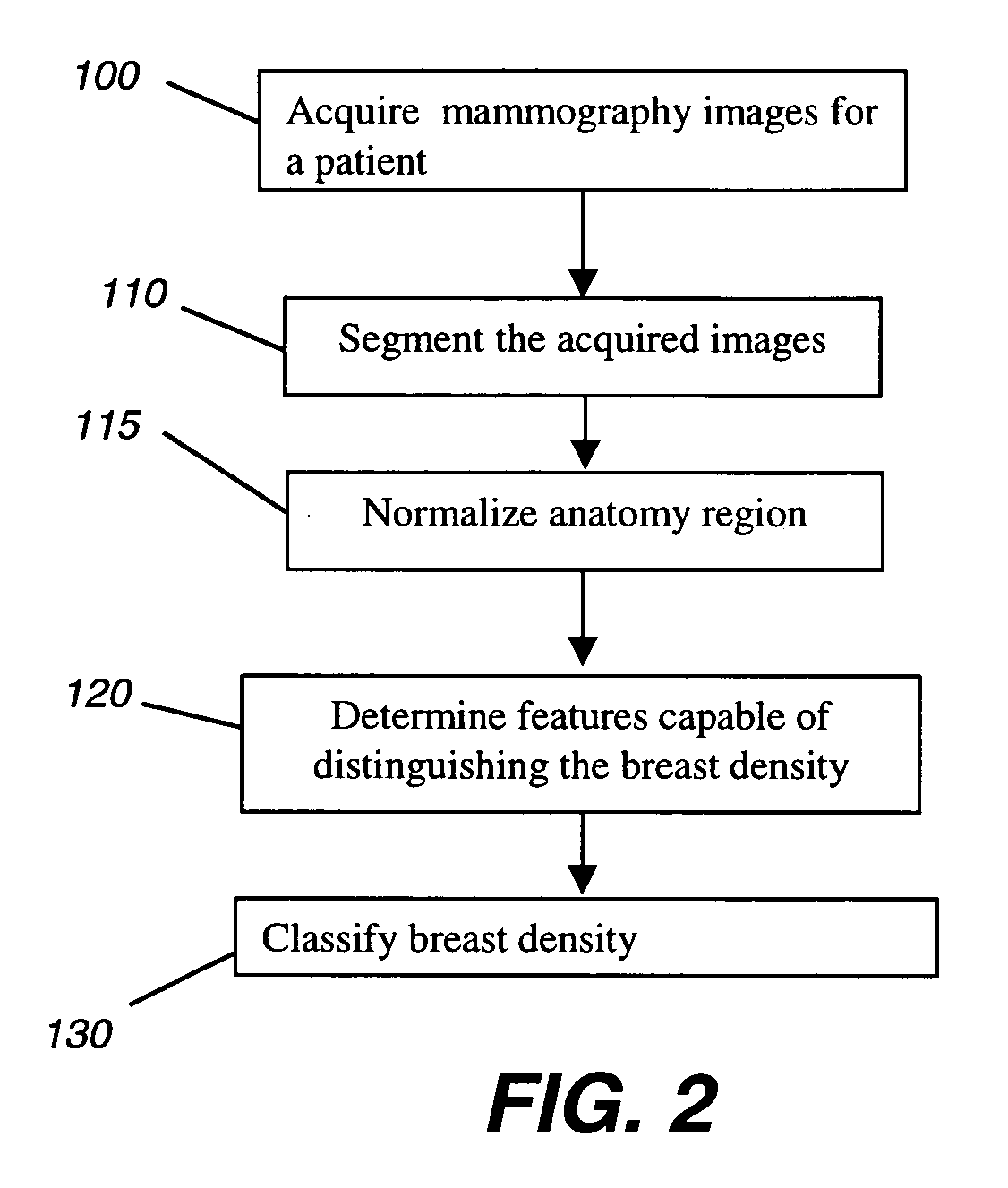
[0024]The present invention is directed to a method for automatically classifying the breast density of mammograms for image rendering and diagnosis. FIG. 2 shows a logic flow generally illustrating an automated method according to the present invention. As shown in FIG. 2, the method includes acquiring / accessing mammogram images of a patient in digital form (step 100); segmenting the breast region from the input mammogram images (step 110); normalizing the anatomy region (step 115); determining features capable of distinguishing the breast density from the segmented breast region (step 120), and classifying the breast density (step 130). These steps will be more particularly describe below.

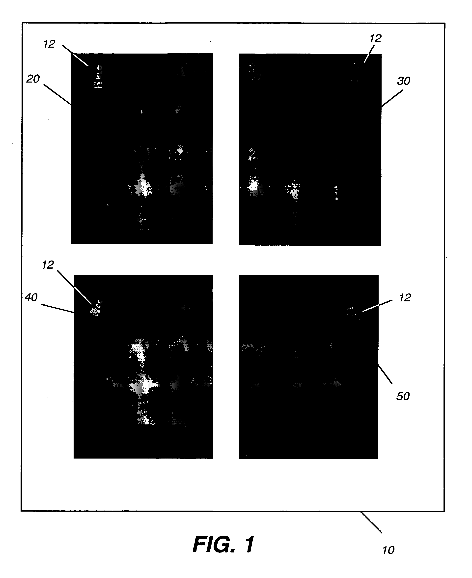
[0025]In image acquisition step 100, mammography images of ...


PUM


Abstract

A method for classifying tissue density of a breast includes obtaining mammography image data and segmenting the mammography image to identify the region representing the breast tissue. A plurality of regions within the breast tissue region are identified for obtaining image features therefrom. A plurality of image features are computed from the identified plurality of regions. The breast tissue density is classified using the computed plurality of image features.

Description

FIELD OF THE INVENTION[0001]The invention relates generally to techniques for processing mammogram images, and in particular, to a method for automatically classifying breast density in mammograms to optimize image rendering and assist diagnosis.BACKGROUND OF THE INVENTION[0002]Screening mammography is a known method for detecting early signs of breast cancer in women. Generally, women undergo an X-ray exam in which X-ray films of the breast are exposed and then developed for review. A radiologist reads the films and assesses the likelihood of the presence of signs of breast cancer. If a suspicious finding is present, the woman will typically be invited for additional, more detailed diagnostic X-ray exams, followed by ultrasonic exams, and possibly biopsy.[0003]In a typical screening exam in the United States of America, four X-rays of the breast are obtained. In conventional practice, two mammographic views are obtained for each breast: a cranio-caudal (CC) view is obtained by posi...

Claims


Application Information

Patent Timeline
IPC IPC(8): G06K9/00
CPCA61B6/502G06T7/0012G06T2207/30068G06T2207/10116G06T7/0083G06T7/12
Inventor LUO, HUIFLETCHER-HEATH, LYNN M.BARSKI, LORI L.
Owner CARESTREAM HEALTH INC
Why Eureka
Try Eureka