Ultrasonic diagnostic device and image processing method

a diagnostic device and ultrasonic technology, applied in the field of ultrasonic diagnostic devices, can solve the problems of deterioration of image quality of three-dimensional images, difficult to perform, in real time, arbitrary image processing for improving image quality, etc., and achieve high speed and satisfactory image quality. , the effect of increasing the load

Inactive Publication Date: 2013-10-17
HITACHI MEDICAL CORP
View PDF5 Cites 10 Cited by
  • Summary
  • Abstract
  • Description
  • Claims
  • Application Information

AI Technical Summary

Benefits of technology

[0022]According to the present invention, it is possible to acquire a three-dimensional image having satisfactory image quality at high speed without increasing a load on an ultrasonic diagnostic device.

Problems solved by technology

However, an ultrasonic image includes a large number of artifacts peculiar to ultrasound such as speckle noise, causing deterioration in image quality of a three-dimensional image.
Since a data amount of three-dimensional data is enormous, it is difficult to perform, in real time, arbitrary image processing for improving the image quality.

Method used


Image

  • Ultrasonic diagnostic device and image processing method
  • Ultrasonic diagnostic device and image processing method
  • Ultrasonic diagnostic device and image processing method

Examples


first embodiment

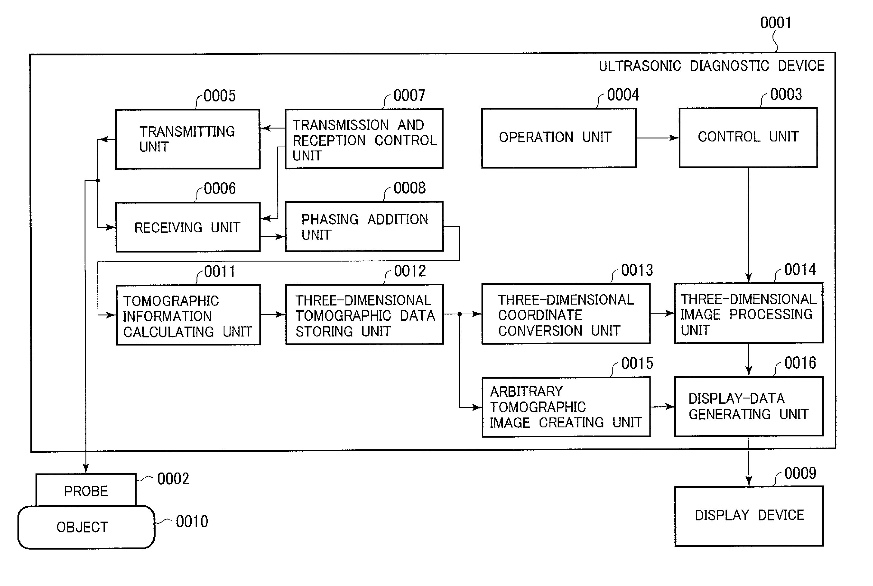
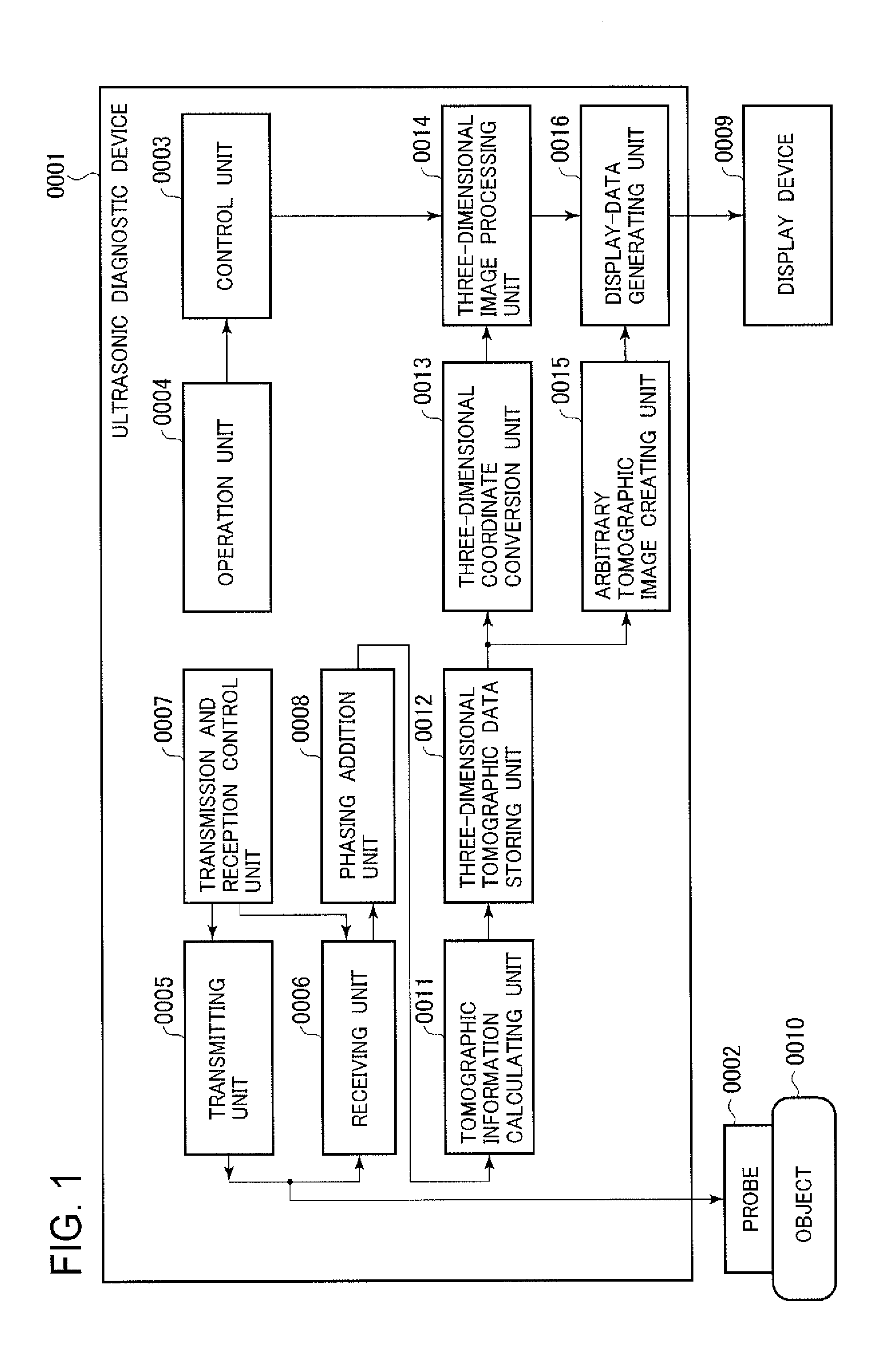
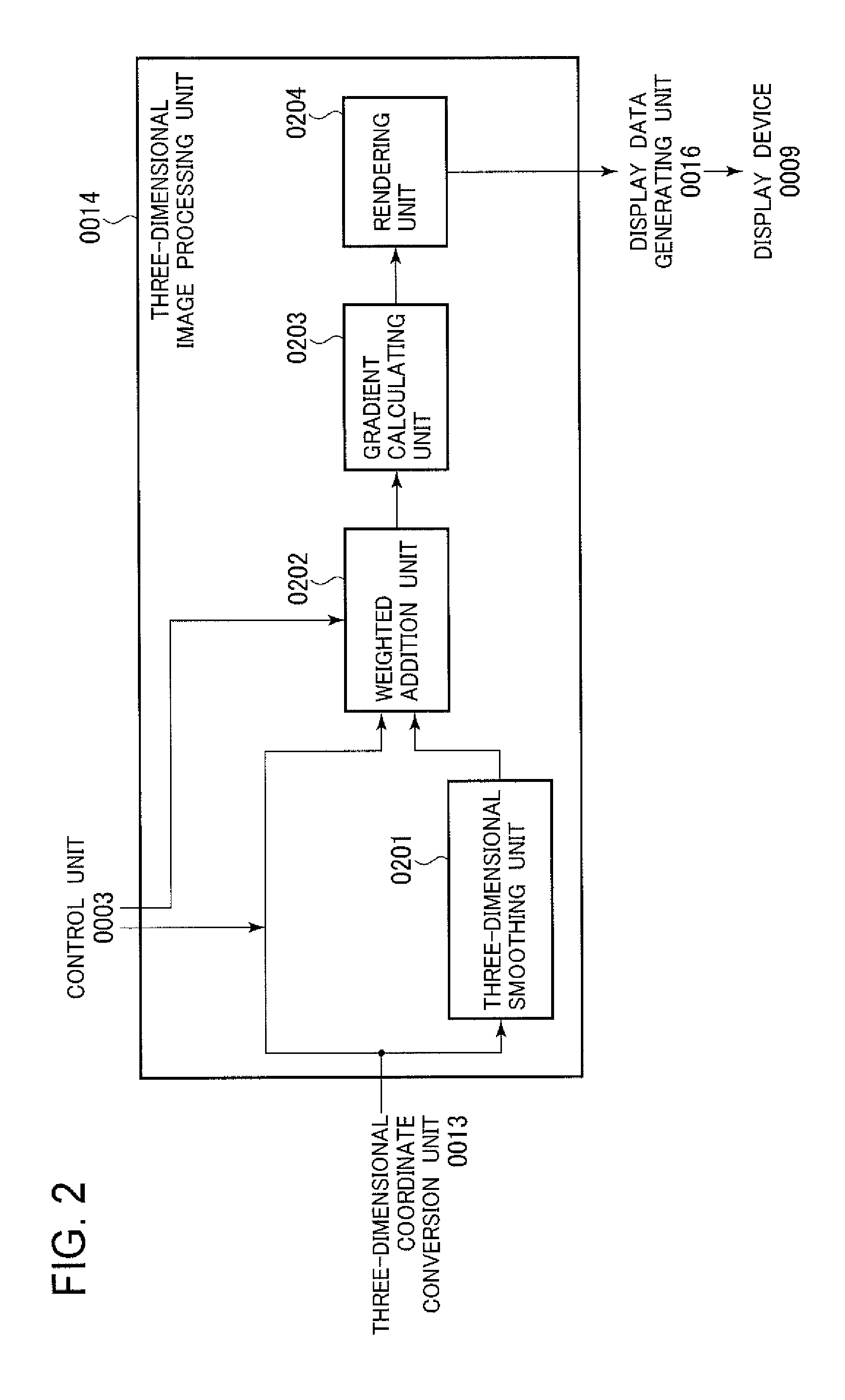
[0038]An embodiment of the present invention is explained below on the basis of the drawings. In the following explanation, in all the figures for explaining the embodiment of the present invention, components having the same functions are denoted by the same reference numerals and signs and repeated explanation of the components is omitted. In this embodiment, a three-dimensional image is generated from combined volume data generated by giving weight set in advance to tomographic volume data after three-dimensional image conversion and smoothed data of the tomographic volume and adding up the data. An ultrasonic diagnostic device includes an ultrasonic probe, a transmitting unit configured to transmit a driving signal to the ultrasonic probe, a three-dimensional tomographic data generating unit configured to generate, using an ultrasonic signal from a test target measured by the ultrasonic probe, tomographic volume data of the test target, a three-dimensional image processing unit ...

second embodiment

[0108]Next, a second embodiment to which the present invention is applied is explained. In the first embodiment, the three-dimensional image processing unit 0014 combines, using a predetermined weight coefficient, tomographic volume data itself, which is an output of the three-dimensional coordinate conversion unit 0013, and volume data obtained by applying the smoothing to the tomographic volume data and generates combined volume data. On the other hand, in this embodiment, a three-dimensional image processing unit adds up, using a predetermined weight coefficient, two volume data respectively obtained by applying different kinds of smoothing to an output of the same three-dimensional coordinate conversion unit and generates combined volume data.

[0109]The ultrasonic diagnostic device 0001 in this embodiment basically has a configuration same as the configuration of the ultrasonic diagnostic device 0001 in the first embodiment. However, since three-dimensional image processing is di...

third embodiment

[0132]Next, a third embodiment to which the present invention is applied is explained. In the second embodiment, the three-dimensional image processing unit 0014a subjects two kinds of volume data, which are obtained by applying the smoothing to an output of the three-dimensional coordinate conversion unit 0013 at different smoothing intensities, to the weighted addition and generates combined volume data. On the other hand, in this embodiment, a three-dimensional image processing unit adds up volume data after a plurality of kinds of filter processing, which are obtained by applying different kinds of filter processing to an output of the same three-dimensional coordinate conversion unit, using a predetermined weight coefficient and generates combined volume data.

[0133]The ultrasonic diagnostic device 0001 in this embodiment basically has a configuration same as the configuration of the ultrasonic diagnostic device 0001 in the first embodiment. However, since the three-dimensional ...


PUM


Abstract

Provided is a technology capable of obtaining at high speed a three-dimensional image having good image quality, without increasing the load on an ultrasonic diagnostic device. A plurality of filter processing having different effects are performed, prior to rendering, on tomographic volume data that has completed coordinate transformation, and volume data is obtained for each. A prescribed weight is added to each voxel for each of the obtained volume data, and a three-dimensional image is generated from the volume data obtained thereby. The filter processing having different effects may be different types of filter processing such as smoothing and sharpening, the same filter processing but having different strengths, or a combination of filter processing and no filter processing.

Description

TECHNICAL FIELD[0001]The present invention relates to an ultrasonic diagnostic device that builds an ultrasonic image of a diagnostic region in an object using ultrasound and displays the ultrasonic image, and more particularly, to an ultrasonic diagnostic device that can build three-dimensional ultrasonic images of a tomographic image, a blood flow image, and elasticity image data.BACKGROUND ART[0002]An ultrasonic diagnostic device transmits ultrasound to an object using an ultrasonic probe, receives a reflected echo signal from the object, and generates a tomographic image of the object using the reflected echo signal. In the ultrasonic diagnostic device, the ultrasonic probe is automatically or manually operated in a minor-axis direction, whereby three-dimensional data is obtained. The obtained three-dimensional data is projected on a two-dimensional projection surface by a method such as volume rendering to display a projected image (a three-dimensional image). For example, bloo...

Claims


Application Information

Patent Timeline
Patent Type & Authority Applications(United States)
IPC IPC(8): G06T15/08
CPCG06T15/08A61B8/0866A61B8/466A61B8/483A61B8/5207A61B8/5246G06T5/004G06T2207/10136G06T2207/30044G01S7/52084G01S15/8993G06T19/00G06T2210/41A61B8/467A61B8/5269
Inventor TSUJITA, TAKEHIRO
Owner HITACHI MEDICAL CORP
Why Eureka
Try Eureka