
X-ray imaging system and image processing device
a technology of image processing and imaging system, applied in the direction of material analysis using wave/particle radiation, image enhancement, instruments, etc., to achieve the effect of low cost and image degradation
- Summary
- Abstract
- Description
- Claims
- Application Information
AI Technical Summary
Benefits of technology
Problems solved by technology
Method used
Image

Examples
Embodiment Construction
[Configuration of X-Ray Imaging System]
[0021]Hereinafter, an embodiment of the present invention is described with reference to the drawings.
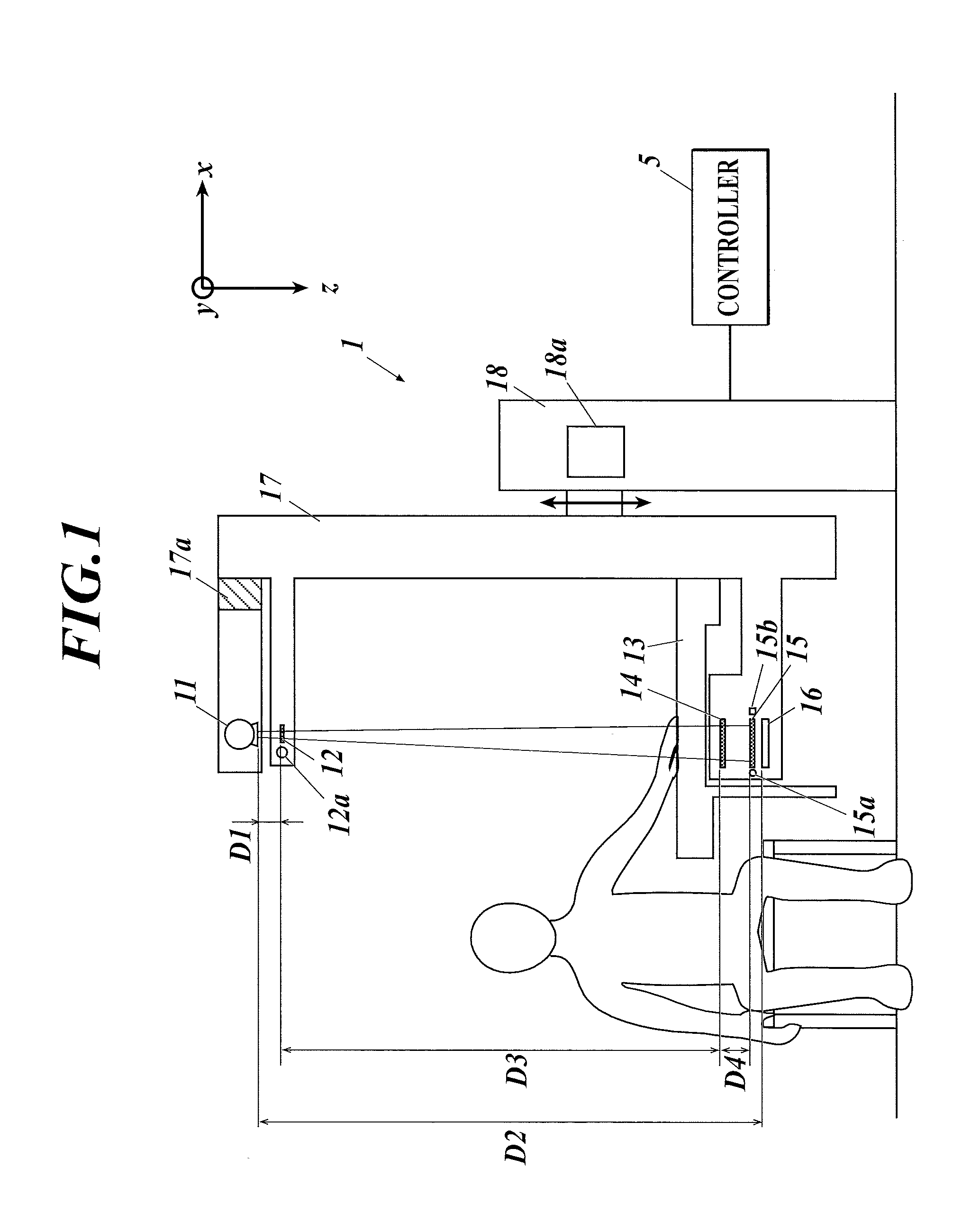
[0022]FIG. 1 shows an example of the configuration of an X-ray imaging system as an embodiment. The X-ray imaging system includes an X-ray imaging device 1 and a controller 5. The X-ray imaging device 1 performs X-ray imaging (fringe scanning) with a Talbot-Lau interferometer, and the controller 5 generates reconstructed images of a subject using a plurality of moire fringe images obtained by the X-ray imaging.
[0023]The X-ray imaging device 1 includes, as shown in FIG. 1, an X-ray source 11, a multi-slit (source grating) 12, a subject table 13, a first grating 14, a second grating 15, an X-ray detector 16, a support unit 17 and a main body unit 18.
[0024]The X-ray imaging device 1 is a vertical type, and the X-ray source 11, the multi-slit 12, the subject table 13, the first grating 14, the second grating 15 and the X-ray detector 16 are arrange...
PUM
| Property | Measurement | Unit |
|---|---|---|
| distance D1 | aaaaa | aaaaa |
| distance D1 | aaaaa | aaaaa |
| height | aaaaa | aaaaa |
Abstract
Description
Claims
Application Information