
Method and biosensor for analysis

a biosensor and analysis method technology, applied in the field of analysis, can solve the problems of high antigen binding, nonspecific binding reduction of biological macromolecules of analytes, etc., and achieve the effect of reducing nonspecific binding and high antigen binding

Inactive Publication Date: 2008-02-19
BEANOR +1
View PDF69 Cites 32 Cited by
  • Summary
  • Abstract
  • Description
  • Claims
  • Application Information

AI Technical Summary

Benefits of technology

[0012]Biomolecule-repellent molecules with suitable terminal anchor groups on a monomer / polymer backbone, or a hydrophilic monomer / polymer “brush” bearing terminal anchor groups can be chemically grafted onto solids like Au, Ag, Cu, Al, Pd and GaAs to form a monolayer, together with biomolecules, especially immunological binder molecules (receptors), such as antibody fragments attached on the same surface. These biomolecule-repellent molecules are directly grafted on the same surface as the biomolecules, especially immunological binder molecules (receptors), such as antibody fragments, so that they form a mixed monolayer. The biomolecule-repellent molecules prevent the nonspecific binding of the analyte in the spaces left between the biomolecules. The immunoreactions can be detected with techniques like surface plasmon resonance, quartz crystal microbalance and impedance. The immobilization procedure will result in a significant reduction of the nonspecific binding of biological macromolecules of the analyte, because the surface between the Fab′-fragments will be highly hydrophilic and non-ionic, and, moreover, a proper orientation of the antibody fragment will result in a high binding of antigen.

Problems solved by technology

The immobilization procedure will result in a significant reduction of the nonspecific binding of biological macromolecules of the analyte, because the surface between the Fab′-fragments will be highly hydrophilic and non-ionic, and, moreover, a proper orientation of the antibody fragment will result in a high binding of antigen.

Method used


Image

  • Method and biosensor for analysis
  • Method and biosensor for analysis
  • Method and biosensor for analysis

Examples


example 1

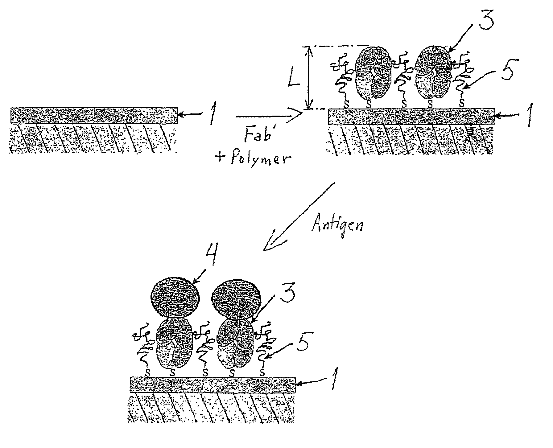
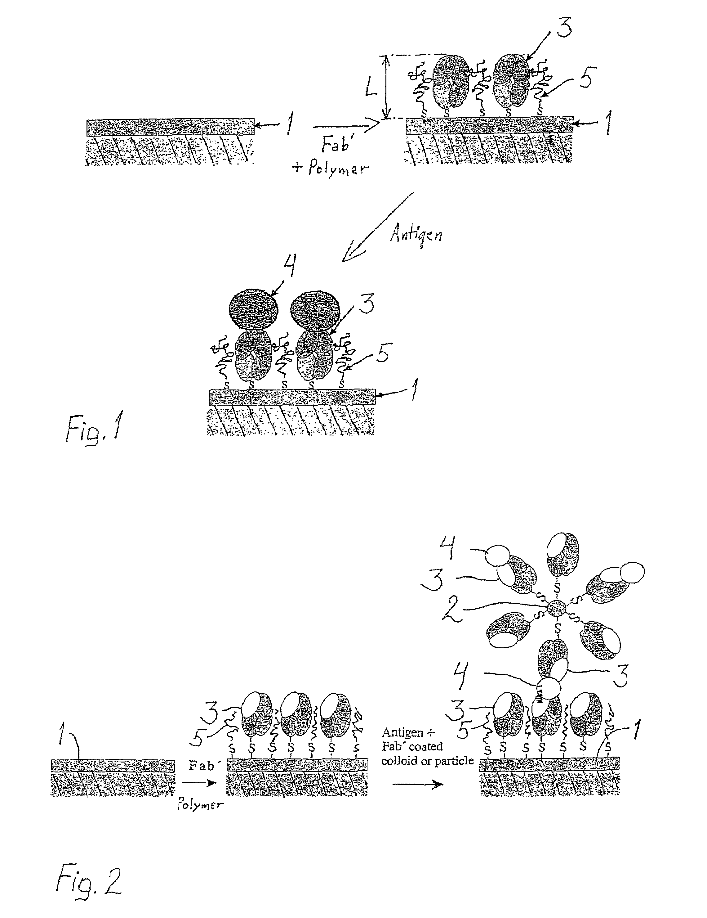
[0040]The model antibody was polyclonal goat anti-human F(ab′)2 (Jackson ImmunoResearch, chromatically purified) with minimized cross-reaction to bovine, horse, and mouse serum proteins. The antigen was chromatically purified human IgG (Jackson ImmunoResearch). F(ab′)2 was split into Fab′-fragments with dithiothreitol (DTT, Merck) prior to use as follows: F(ab′)2 with a concentration of 1.2-1.3 mg / ml (100 μl) was mixed with 50 μl of HEPES / EDTA buffer (150 mM NaCl, 10 mM HEPES, 5 mM EDTA, pH=6.0) and 10 μl of a 0.1 M DTT solution in HEPES / EDTA buffer in a microdialysis tube. The dialysis tube was immersed in 250 ml of argon-purged HEPES / EDTA buffer and dialyzed for about 18 h at room temperature under argon. The Fab′-fragment was maintained under argon and used immediately for attachment.

[0041]Fab′-fragments have well-accessible reactive thiol groups opposite the antibody binding domains.

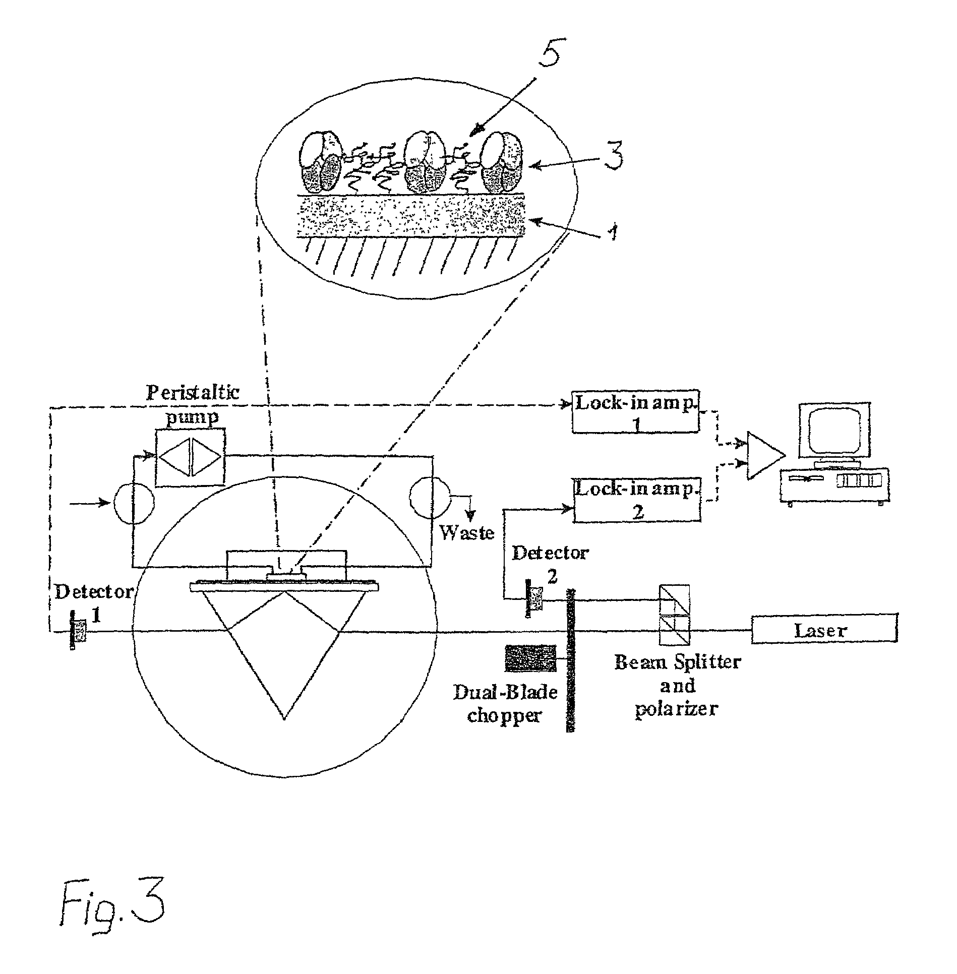
[0042]A Surface Plasmon Resonance device, SPRDEVI was used for the measurements (VTT, Tampere). T...


PUM

PropertyMeasurementUnit
wavelengthaaaaaaaaaa
pHaaaaaaaaaa
concentrationaaaaaaaaaa

Abstract

In a method for analysis biomolecules (3) attached to a solid surface of a substrate (1) are used for detecting the presence of analytes (4) in a sample by binding of the analytes to the biomolecules. The biomolecules (3) are attached directly to the surface of the substrate together with biomolecule-repellent molecules (5), which cover the surface between the biomolecules (3) to prevent nonspecific binding of analytes (4) and other biomolecules. The invention relates also to a biosensor where biomolecules (3) are attached directly to the substrate (1) together with biomolecule-repellent molecules (5), which cover the surface between the biomolecules (3) to prevent non-specific binding of analytes (4) and other biomolecules. The biomolecules (5) can be self-assembled hydrophilic polymers. One example of using the invention is immunological analysis using surface plasmon resonance (SPR).

Description

FIELD OF THE INVENTION[0001]The present invention relates to a method for analysis. The invention also relates to a biosensor for carrying out the method.BACKGROUND OF THE INVENTION[0002]There is an increasing need for simple, rapid and easy-to-use sensors to aid clinical diagnosis and for environmental analysis.[0003]The very specific and tight binding between antibodies and antigens has traditionally been measured in clinical laboratories by radioimmunoassay, solid phase enzymeimmunoassay and fluoroimmunoassay. Quantification of the antibody-antigen complex relies on a marker molecule, such as a radioisotope, an enzyme or a fluorescent probe. In most cases, the result is not obtained until several incubations, washing and separation steps have been performed.[0004]Presently, immunosensors that can monitor antigen concentrations in real-time with the aid of an immobilized antibody and that do not require the use of labeled species are being developed. The lack of control over the o...

Claims


Application Information

Patent Timeline
Patent Type & Authority Patents(United States)
IPC IPC(8): C12M1/34G01N5/02G01N21/27G01N33/543
CPCG01N33/54373G01N33/54393Y10S435/808Y10S435/962Y10S436/81Y10S436/805Y10S436/806
Inventor VIKHOLM, INGERSADOWSKI, JANUSZ
Owner BEANOR
Learn More