Method and medical imaging apparatus for planning an image acquisition based on a previously-generated reference image

a technology of medical imaging and reference image, applied in the field of planning an examination, can solve the problem that the post-processing procedures of individual images cannot be reconstructed in the prior art, and achieve the effect of accelerating the planning of examination

Inactive Publication Date: 2010-05-25
SIEMENS HEALTHCARE GMBH
View PDF18 Cites 64 Cited by
  • Summary
  • Abstract
  • Description
  • Claims
  • Application Information

AI Technical Summary

Benefits of technology

[0009]An object of the present invention is to simplify and to accelerate the planning of examinations so that among other things, reference images for the generation of which a number of initial images and / or a number of post-processing steps were necessary, can be used in a simple manner.

Problems solved by technology

Only individual measurements or their parameters could be incorporated into the workflow planning in the prior art, but the possibly-present post-processing procedures of the individual images could not be reconstructed in the prior art.

Method used


Image

  • Method and medical imaging apparatus for planning an image acquisition based on a previously-generated reference image
  • Method and medical imaging apparatus for planning an image acquisition based on a previously-generated reference image
  • Method and medical imaging apparatus for planning an image acquisition based on a previously-generated reference image

Examples


Embodiment Construction

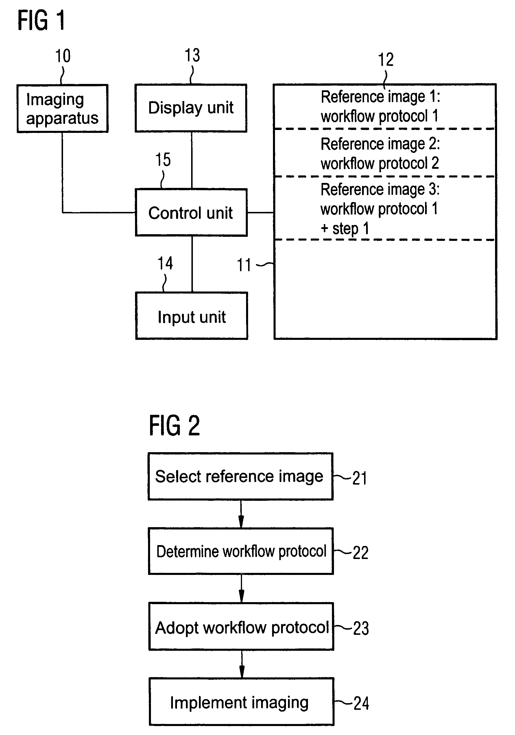
[0022]An imaging diagnosis device (here a magnetic resonance system) is schematically shown in FIG. 1. The functioning of a magnetic resonance system for generation of slice images of the body by nuclear magnetic resonance is known to those skilled in the art and need not be described in detail herein. For clarity, only the components that are necessary for understanding of the present invention are shown.

[0023]The magnetic resonance system has an imaging apparatus (scanner) that, as is known in the prior art, acquires MR images of the examination subject. In the planning of a new examination, the operating person of the magnetic resonance system can access reference images stored in a storage unit 11, when images of the current examination person are to be generated that correspond to the reference image with the respective image acquisition parameters. Data sets 12 are stored in the storage unit 11. A data set 12 includes a reference image and the associated workflow protocol that...


PUM


Abstract

In a method and device for planning an examination of an examination subject in an imaging diagnosis device, a reference image is selected, and an image of the examination subject is to be made that corresponds to the reference image. A workflow protocol is determined with which the reference image was generated. The acquisition of all required images of the examination subject is planned using this workflow protocol. The workflow protocol is executed for creation of an image of the examination subject that corresponds to the reference image.

Description

BACKGROUND OF THE INVENTION[0001]1. Field of the Invention[0002]The present invention concerns a method for planning an examination of an examination subject in an imaging diagnosis device as well as an imaging diagnosis device for this purpose. The invention can in particular (but not exclusively) be applied in magnetic resonance systems in which the magnetic resonance images (MR images) that were acquired by the examination subject can be post-processed in different manners.[0003]2. Description of the Prior Art[0004]Magnetic resonance systems are increasingly used in radiology in the examination of patients since they enable a relatively exposure-free examination of the patient as well as the appraisal of different body regions with different contrast performance. Many different post-processing possibilities have been developed in order to be able to process the acquired MR images with various post-processing steps and to be able to better respond to a specific clinical question.[...

Claims


Application Information

Patent Timeline
Patent Type & Authority Patents(United States)
IPC IPC(8): A61B5/05G16H30/20G16H30/40
CPCA61B5/055G06F19/3406G06F19/321G16H40/63G16H30/20G16H30/40
Inventor BECK, WALTERMAYER, KLAUSMOHR, CECILEZELTNER, JOCHEN
Owner SIEMENS HEALTHCARE GMBH
Why Eureka
Try Eureka