
Method device and system for monitoring lung ventilation

a technology for monitoring devices and lung ventilation, applied in the field of lung ventilation, can solve problems such as inability to fix the inner end, misplacement of the tube, both during intubation or during ventilation, and can have dire consequences, and achieve the effects of reducing the number of patients

Active Publication Date: 2012-07-24
TECHNION RES & DEV FOUND LTD
View PDF6 Cites 15 Cited by
  • Summary
  • Abstract
  • Description
  • Claims
  • Application Information

AI Technical Summary

Problems solved by technology

In small children and neonates there is no cuff and it is impossible to fix the inner end.
It is recognized that the position of the tube within the trachea is of utmost importance because such tubes are necessary to ensure that a patient's lungs receive oxygen, and any misplacement of the tube, both during the intubation or during ventilation, can have dire consequences.
Asymmetric or one lung ventilation may lead to the development of respiratory insufficiency and subsequent hypoxia, hypercapnia and acidosis.
Furthermore, one lung inflation can cause hyperinflation of one side leading to pneumothorax at that side, and simultaneously can create atelectasis at the other side, leading to other complications as lobar pneumonia, and the like.
Conversely, displacement of the tube to or above the vocal cords may result in insufficient ventilation and / or damage to the vocal cord.
Such barotraumas are caused by lung disease and / or high air pressure in the lungs which results in rupture of alveolar structures and lung tissue.
Another complication which may occur during ventilation is deterioration in the gas supply, due to, e.g., malfunction of the mechanical ventilator, partial detachment of ventilator circuit, partial obstruction in the endotracheal tube by secretions, fluids accumulation, torsion, kink, and the like.
Of particular challenge is the ventilation of neonate and premature infants, because the endotracheal tube is extremely small and any small displacement (within millimeters) can result in inappropriate ventilation.
Serious life threatening complications can result from inappropriate ventilation.
A most dangerous complication is partial or full detachment of the endotracheal tube with ineffective ventilation.
The associated hypoxia and hemodynamic changes induced are especially dangerous for the vulnerable premature newborn infant and can lead to intracranial (intraventricular) hemorrhage and to the development of severe irreversible neurological sequelae as a result of brain damage.
Traditional monitoring techniques, however, suffer from many limitations, including non-automated monitoring which requires tight physical supervision of the medical staff, frequent false alarms events, slow speed of detection, low sensitivity, particularly with thin tubes, low specificity and low sensitivity, particularly to the detection of pneumothorax or other complications associated with asymmetric ventilation.

Method used


Image

  • Method device and system for monitoring lung ventilation
  • Method device and system for monitoring lung ventilation
  • Method device and system for monitoring lung ventilation

Examples


example 1

Ventilation Indices

[0144]Following is a list of ventilation indices which can be used to characterize the ventilation status, according to various exemplary embodiments of the present invention. Each of the following ventilation indices is preferably calculated as a function of time and for each sensing location. An averaging procedure can also be employed either over a predetermined period of time and / or over two or more sensing locations. Additionally, an interpolation procedure can be employed so as to obtain spatial distribution of the ventilation index. Thus, each of the following ventilation indices can be a function of both time and space, a function of time, a function of space or a number.

Tidal Motion Index (TMI)

[0145]The TMI is preferably defined to describe the local peak-to-peak amplitude changes of the chest wall displacement during ventilation. The TMI can be calculated by subtracting the lowest amplitude of the chest wall from the highest amplitude of the chest wall o...

example 2

Ventilated Lung Model

[0155]A simplified scheme of a ventilated lung is illustrated in FIG. 4a.

[0156]Vin represents the mechanical ventilation machine generating a pulsatile pressure or flow.

[0157]R_T represents the impedance to flow through the endotracheal tube. The thin tubes (3-4 millimeters in diameter for neonates) impose high resistance, especially at high velocity.

[0158]R_L represents the air leak through the upper air ways (mouth and nose), especially when the tip of the tube in place in the oropharynx. It is described by a variable resistor. When the tube is displaced to the oropharynx this resistance decreases to simulate the large leak. When the tube is placed in the appropriate position and a balloon is inflated to fix the position of the tube and to prevent leak this resistance approach infinity.

[0159]R_SS, R_SP and C_S represent the components of the impedance to flow into the stomach. This impedance include a serial resistive element (flow in the esophagus) R_SS, and...

example 3

Experimental Trial

[0166]Following is a description of an experiment in which chest wall motion in a ventilated rabbit model was monitored according to preferred embodiments of the present invention using a prototype system.

Methods

[0167]The experiments were performed according to the guidelines approved by the Institutional Animal Experimental Ethics Committee of the Technion-Israeli Institute of Technology. The experimental study was carried in 7 adult male rabbits, 2.3±0.2 Kg of weight. The experimental subjects were anesthetized by intramuscular injection of Xylazine (10 mg / kg), Ketamine (90 mg / kg) and Fentanyl (0.2-0.6 ml / kg) with repeated doses during the experiments. Endotracheal intubation was performed by direct laryngoscopy and a 3 mm diameter endotracheal tube (ETT—Portex Ltd., UK) was introduced into the trachea.

[0168]Mechanical ventilation was initiated and ventilatory parameters were standardized in order to keep the blood PH, PO2, and PCO2 within physiological limits. E...


PUM


Abstract

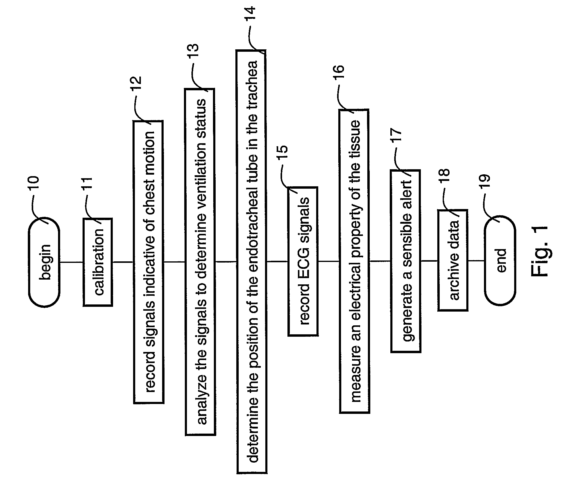
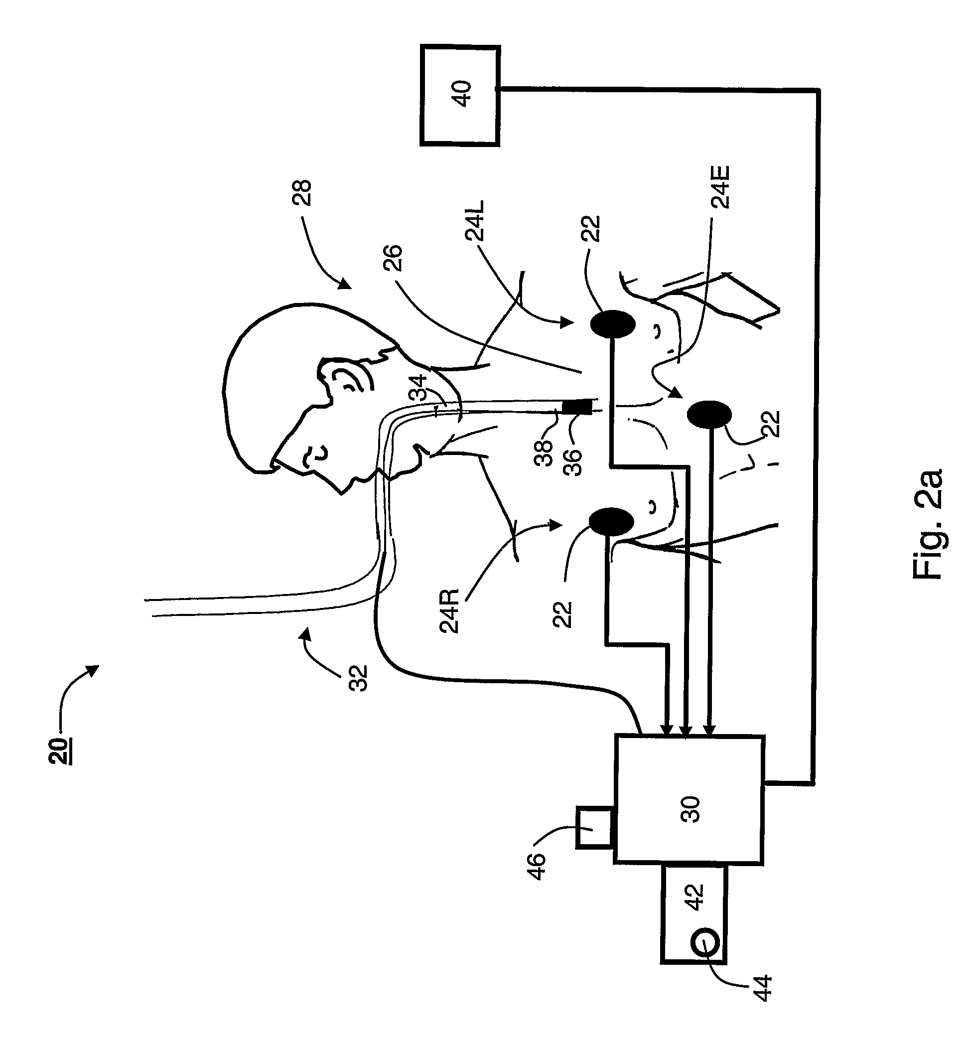
A method of monitoring lung ventilation of a subject is disclosed. The method comprises recording signals from a plurality of sensing location on the chest of the subject, at least a portion of the signals being indicative of a local motion of the chest at a respective sensing location. The method further comprises operating a data processing system to analyze the signals such as to determine a status of the ventilation, thereby to monitor the lung ventilation of the subject.

Description

FIELD AND BACKGROUND OF THE INVENTION[0001]The present invention relates to lung ventilation and, more particularly, to a method device and system suitable for monitoring lung ventilation.[0002]In the medical treatment of patients requiring breathing assistance, it is common to insert an endotracheal or nasotracheal tube into the trachea of the patient, by way of the mouth, nose or any other surgically created opening. One end of the tube is connected to a ventilator which periodically forces air into the lungs through the tube. In big children and adult patients the inner end of the tube is typically provided with an inflatable cuff which is inflated by conventional means subsequently to the insertion of the tube into the trachea. In small children and neonates there is no cuff and it is impossible to fix the inner end.[0003]It is recognized that the position of the tube within the trachea is of utmost importance because such tubes are necessary to ensure that a patient's lungs rec...

Claims


Application Information

Patent Timeline
Patent Type & Authority Patents(United States)
IPC IPC(8): A61B5/08A61B5/087
CPCA61B5/08A61B5/1135A61M16/0057A61B5/0205A61B5/4233A61M16/0051A61B5/0402A61B5/06A61B5/091A61B2562/0219A61B5/062A61M16/021A61B5/318A61M16/0069A61M16/04A61M2205/15A61M2205/3303A61M2210/105A61M2230/04A61M2230/40A61M2230/63A61M2240/00A61M2250/00
Inventor LANDESBERG, AMIRWAISMAN, DANLEVY, CARMIT
Owner TECHNION RES & DEV FOUND LTD
Learn More