Supercharge Your Innovation With Domain-Expert AI Agents!

Perfusion assessment based on animated perfusion imaging

A medical imaging system and image input technology, which is applied in the display field of animated perfusion images, can solve problems such as not allowing direct intuitive perception of the perfusion process

Active Publication Date: 2010-01-27
BRACCO SUISSE SA
View PDF6 Cites 0 Cited by
  • Summary
  • Abstract
  • Description
  • Claims
  • Application Information

AI Technical Summary

Problems solved by technology

[0010] However, parametric images only provide a static representation of perfusion parameter values
Therefore, this does not allow for a direct intuitive perception of the perfusion process, which is usually provided by playback of the original image sequence

Method used


Image

  • Perfusion assessment based on animated perfusion imaging
  • Perfusion assessment based on animated perfusion imaging
  • Perfusion assessment based on animated perfusion imaging

Examples


Embodiment Construction

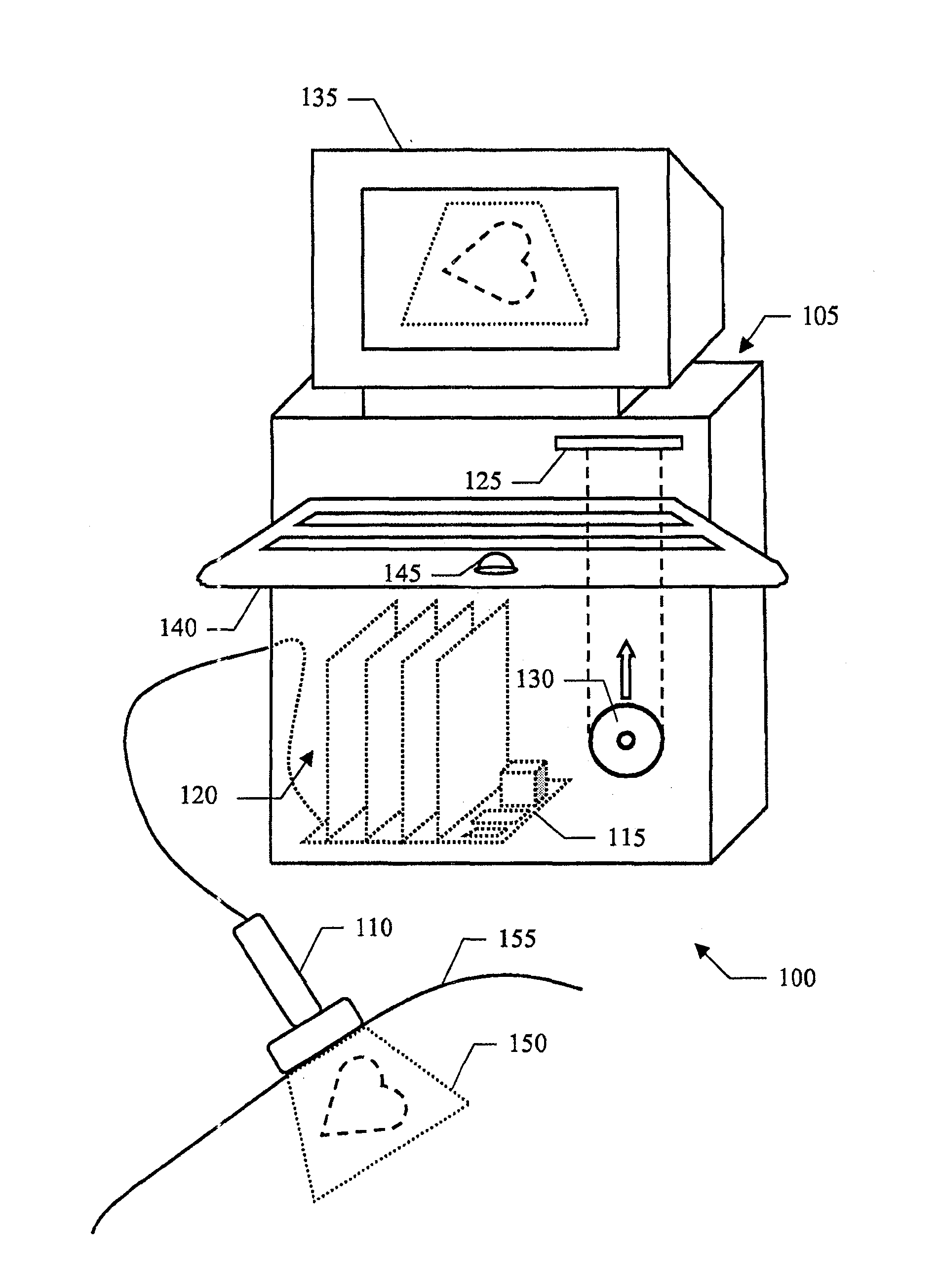
[0046] specifically refer to figure 1 , illustrating a medical imaging system 100 . In particular, the system 100 includes an ultrasound scanner with a central unit 105 of a hand-held transmit-receive imaging probe 110 (eg, of the matrix array type). The imaging probe 110 transmits ultrasound waves comprising a pulse sequence (for example, having a center frequency of 2-10 MHz), and receives radio frequency (RF) echo signals generated by backscattering of the ultrasound waves; for this purpose, the imaging probe 110 is provided with a The pulse-echo mode uses the transmit / receive multiplexer of the imaging probe 110 .

[0047] The central unit 105 includes a motherboard 115 on which electronic circuits controlling the operation of the ultrasound scanner 100, such as a microprocessor, working memory, and a hard disk drive, are mounted. Also, one or more daughter boards (indicated generally at 120) plug onto main board 115; daughter boards 120 provide the electronics that driv...


PUM


Abstract

A medical imaging system (100) is proposed. The system includes means (203- 212) for providing a sequence of recorded input images each one offering a digital representation at a corresponding instant of a body part being perfused with a contrast agent, each input image including a plurality of visualizing values each one representing a corresponding portion of the body part, and means (214-218) for associating each sequence of corresponding sets in the input images of at least one visualizing value with a model function of time; the system further includes means (225-233;705-740) for generating a sequence of computed images at further instants, each computed image including a plurality of further visualizing values each one being determined by an instantaneous function-value which is calculated from the associated model function at the corresponding further instant, and means (239-242) for displaying the sequence of computed images.

Description

technical field [0001] The invention relates to the field of medical imaging. More particularly, the invention relates to the display of animated perfusion images. Background technique [0002] Medical imaging is a well-established technology in the field of equipment for medical applications. In particular, this technique is commonly used in the evaluation of blood perfusion, which finds use in several diagnostic applications, especially in ultrasound analysis. For this purpose, an ultrasound contrast agent (UCA) such as a suspension comprising phospholipid-stabilized gas-filled microbubbles is injected into the patient. The contrast agent acts as an effective ultrasound reflector so that it can be easily detected by applying ultrasound and recording the resulting echo signal. As the contrast agent flows at the same speed as the blood in the patient, its traces provide information about blood perfusion in the analyzed body part. [0003] Typically, the flow of contrast ...

Claims


Application Information

Patent Timeline
Patent Type & Authority Patents(China)
IPC IPC(8): A61B8/00A61B8/06G06T7/00G01S15/89
CPCA61B8/461G06T2207/30004A61B8/13A61B8/463G06T7/20A61B8/481G01S15/8977A61B8/5238A61B8/06
Inventor 玛索·阿迪特尼古拉斯·罗格宁彼得·弗林京
Owner BRACCO SUISSE SA
Features
  • R&D
  • Intellectual Property
  • Life Sciences
  • Materials
  • Tech Scout
  • Unparalleled Data Quality
  • Higher Quality Content
  • 60% Fewer Hallucinations
Learn More