Use of a topical composition containing epidermal growth factor (EGF) for diabetic foot amputation prevention

An epidermal growth factor, topical technology, used in medical preparations containing active ingredients, drug combinations, skin diseases, etc.

Inactive Publication Date: 2009-02-25
CENT DE ING GENETICA & BIOTECNOLOGIA
View PDF2 Cites 3 Cited by
  • Summary
  • Abstract
  • Description
  • Claims
  • Application Information

AI Technical Summary

Problems solved by technology

Furthermore, because the tissues of those types of diabetic foot lesions contain much higher levels of proteolytic enzymes than normal skin (Yager D.R., Chen S.M., Ward S.I. et al., (1997) Ability of chronic wound fluids to degrade peptide growth factors is associated withinincreased levels of enastase activity and diminished levels of proteinase inhibitors.Wound Rep.Reg.5:23-32), the active pharmaceutical composition may be enzymatically degraded to a greater extent

Method used


Image

  • Use of a topical composition containing epidermal growth factor (EGF) for diabetic foot amputation prevention
  • Use of a topical composition containing epidermal growth factor (EGF) for diabetic foot amputation prevention
  • Use of a topical composition containing epidermal growth factor (EGF) for diabetic foot amputation prevention

Examples


Embodiment 1

[0021] Example 1. Production of EGF-loaded liposomes

[0022] Phosphatidylcholine was dissolved at a concentration of 10 mg / mL in absolute ethanol in a 50 mL round bottom flask. Solvent was removed by rotary evaporation until a dry lipid film formed on the flask walls. For encapsulation of EGF in liposomes, the dried lipid film was hydrated with a buffered aqueous solution containing EGF and homogenized by stirring. To reduce the size of the vesicles, the suspension was subjected to several extrusions through a 100 nm pore size polycarbonate membrane until the average size of the vesicles was about 100 nm. The EGF-loaded liposome suspension was centrifuged at 100 000 x g for 40 min at 4°C to separate non-encapsulated EGF from encapsulated EGF. The supernatant was transferred to a new tube and the pellet was resuspended in pH 7.2 phosphate buffered saline. The centrifugation step was repeated again under the same conditions and the supernatant was transferred to a new tube a...

Embodiment 2

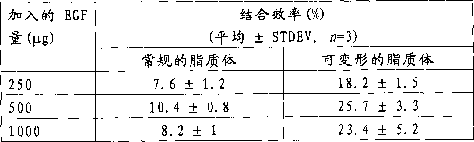
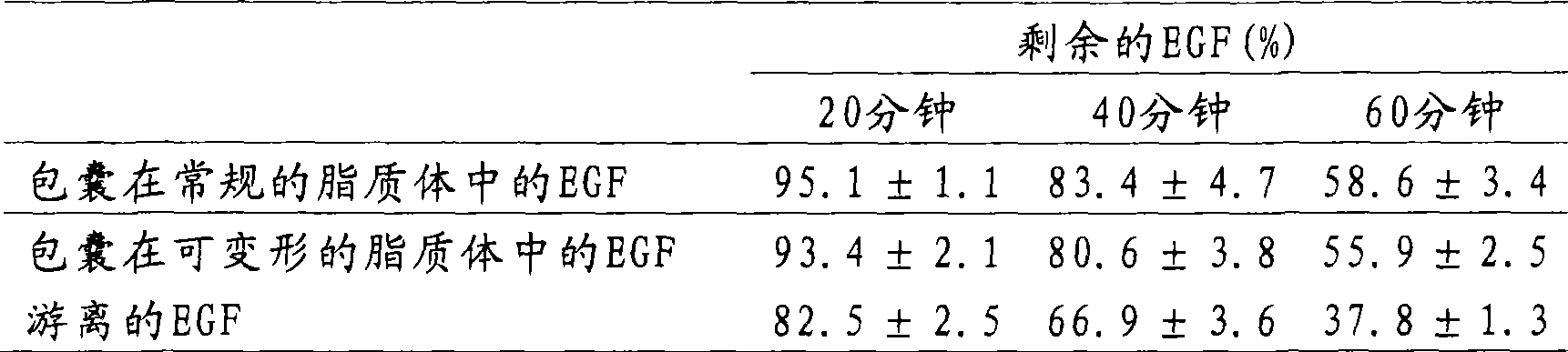
[0025] Embodiment 2. Determination of the encapsulation efficiency of EGF in liposome

[0026] To determine the incorporation efficiency of EGF in liposomes, liposome suspensions were high-speed centrifuged and the protein content of the resulting pellet (EGF-loaded liposomes) and supernatant (free EGF) was determined. The liposome suspension was centrifuged at 100 000 x g for 40 min at 4 °C. The supernatant was transferred to a new tube and the pellet was resuspended in pH 7.2 phosphate buffered saline. The centrifugation step was repeated again under the same conditions and the supernatant was transferred to a new tube and mixed with the supernatant of the first centrifugation step. The pellet (comprising EGF-loaded liposomes) was resuspended in 500 μL of pH 7.2 phosphate-buffered saline. Then, the protein of the pellet or supernatant was extracted and separated from the lipid by adding 0.5% (v / v) Triton X-100 to the sample, and they were subjected to reverse phase high pe...

Embodiment 3

[0029] Example 3. Determination of liposome size and morphology

[0030] Liposome samples were analyzed by transmission electron microscopy to determine liposome size and morphology. Liposomes were visualized by negative staining with uranyl acetate. Negatively stained samples were observed under a transmission electron microscope Jeol-JEM 2000EX operated at 80KV. Electron micrographs corresponding to each liposome preparation were digitized using a scanner and the diameter of the liposomes was measured using DIGIPATVersion 3.3 software (EICSOFT, Havana, Cuba). Particle size is the average value of all liposomes present in the respective micrographs and is expressed as the mean ± standard deviation of three individual determinations.

[0031] Preparations of deformable or conventional liposomes loaded with EGF consisted of homogeneous populations of spherical or oval vesicles. The average size of the vesicles was 130±7 nm and 123±4 nm for regular and deformable liposomes, r...


PUM


Abstract

The invention relates to the use of topical formulations containing epidermal growth factor which is encapsulated or associated with standard or deformable liposomes, which are intended to be applied to and around chronic ischemic skin lesions. The inventive formulations, in contrast to the prior art, are useful in that they enable a high bioavailability of the epidermal growth factor in the deep tissue below the lesion and for preventing diabetic foot amputation.

Description

field of invention [0001] The present invention relates to topical formulations comprising epidermal growth factor (EGF) encapsulated in or associated with deformable or conventional liposomes for administration in chronic ischemic On and around the surface of skin lesions for the prevention of diabetic amputations. Background of the invention [0002] Diabetes mellitus and its complications are the leading non-traumatic causes of lower extremity amputations. This is a medical challenge of increasing importance as the incidence and incidence of diabetes will increase due to population aging and sedentary lifestyles. At least 15% of diabetic patients develop chronic ulcers on both feet during their lifetime, and it is estimated that 20% of these patients require lower extremity amputations (Reiber G.E., Boyko E.J., Smith D.G. (1995) Lower extremity footulcers and amputations in diabetes. in: Harris M.I., Cowie C.C., Reiber G., Boyko E., Stern M., Bennett P. eds. Diabetis in...

Claims


Application Information

Patent Timeline
Patent Type & Authority Applications(China)
IPC IPC(8): A61K38/18A61K9/127A61P17/02
CPCA61K9/127A61K38/1808A61P17/00A61P17/02A61P3/10A61P43/00A61P9/00A61K38/18
Inventor B·Y·贝坦克特罗德里格斯E·普波埃斯卡罗纳R·派斯梅雷莱斯J·A·贝兰加阿科斯塔
Owner CENT DE ING GENETICA & BIOTECNOLOGIA
Why Eureka
Try Eureka