II type collagen sponge bracket and uses thereof

A collagen sponge and collagen technology, applied in the field of biomedicine, can solve the problems of being difficult to use as a cell culture scaffold material, lack of mechanical strength, and difficult to achieve cartilage repair, etc., and achieve a good repair effect

Inactive Publication Date: 2009-10-07
广州市红十字会医院
View PDF1 Cites 18 Cited by
  • Summary
  • Abstract
  • Description
  • Claims
  • Application Information

AI Technical Summary

Benefits of technology

This patented technology describes how it works well when applied on damaged or degenerated joint surfaces such as knee menisci (the main symptom being addressed). It was found that this material could break down over time within two weeks after surgery compared with older methods like autograft transplantation.

Problems solved by technology

The technical problem addressed in this patented text relates to finding suitable sources of type-2 collagen for use in making prosthetic joint repairs without causing inflammatory responses during surgery. Current options involve complicated processes involving multiple steps like physical removal of damaged parts followed by chemical agents or enzymes, leading to potential complications including bleeding, necrosis, fibrocyst growth, poor integration between arthritis/osteoarthropathy patients' own skeletal muscle, reduced fertility rates caused by excessive calcium intake compared to those who donate their own ones.

Method used


Image

  • II type collagen sponge bracket and uses thereof
  • II type collagen sponge bracket and uses thereof
  • II type collagen sponge bracket and uses thereof

Examples


Embodiment 1

[0031] 1. Preparation of high-purity type II collagen [Reference: Li Siming, Ye Chunting, etc., Preparation and detection of high-purity porcine cartilage type II collagen, Journal of Biomedical Engineering, 2001, 18(4)592-594]:

[0032] Material:

[0033] Fresh pig articular cartilage: obtained from the limb articular cartilage of freshly slaughtered pigs in the slaughterhouse

[0034] Guanidine hydrochloride: Farco 500g / bottle

[0035] Pepsin: Sigma 100g / bottle

[0036] The rest are domestic analytical reagents.

[0037] method:

[0038] Cut the hyaline cartilage from freshly slaughtered pig limb joints, cut into thin slices, degrease, mash, and homogenate, then stir with 10 times the volume of 4M (pH7.5) guanidine hydrochloride for 24 hours, and centrifuge; the sediment diameter is fully washed , Weigh the precipitated cartilage particles with a wet weight of about 260g, use pepsin with a mass ratio of 1 / 50 to enzymolyze under acidic conditions for 24-48h, centrifuge, a...

experiment example 1 II

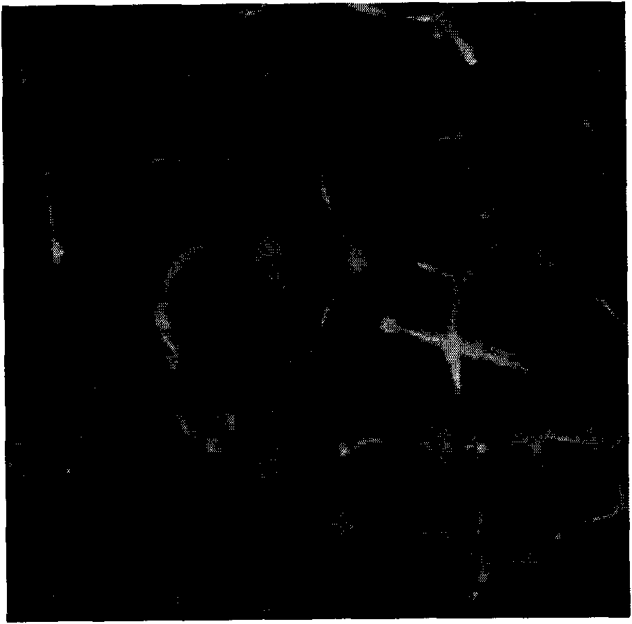
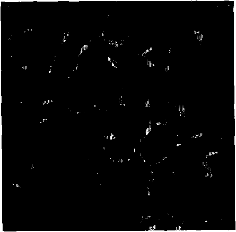
[0044] Experimental example 1. Pore diameter detection of type II collagen sponge scaffold material

[0045] Type II collagen sponge materials were prepared according to the above method, and were divided into stock solution non-crosslinking group, stock solution crosslinking group, concentrated non-crosslinking group, and concentrated crosslinking group.

[0046] Collagen autofluorescence layered scanning of collagen sponges with various pore diameters was carried out by laser confocal microscope. The excitation wavelength was 488nm, and the emission wavelength was 520nm. The autofluorescence signal intensity was measured to determine the pore size of the sponge material; using CLSM image The analysis system is used for operation and analysis, and then the SPSS13.0 software package is used to make statistics on the data.

[0047] As shown in Figure 2, the laser confocal microscope results show that the collagen sponges of each group have obvious autofluorescence, indicating t...

experiment example 2

[0052] Experimental Example 2: Solubility Detection of Type II Collagen Scaffold Materials

[0053] The type II collagen sponge material was prepared according to the above method, which was divided into a crosslinked group and an uncrosslinked group.

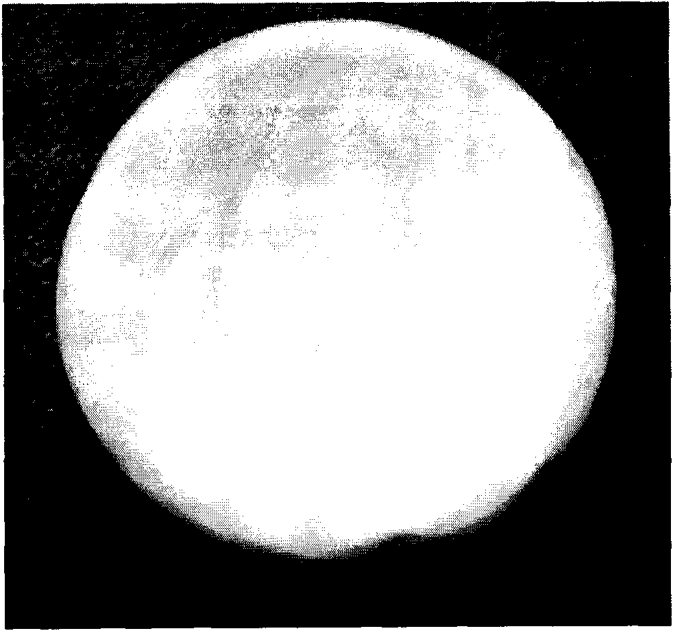
[0054] Cut the type II collagen sponge material into 1×1cm 2 Put it into a 6-well culture plate, add 3ml of DMEM medium, and observe the condition of the sponge at 37°C.

[0055] The results showed that the non-cross-linked type II collagen sponge material swelled after being added to the culture medium, and partially dissolved, and the material became soft and translucent; after 7 days, the material was completely dissolved. The appearance of the cross-linked sponge material remains unchanged after being added to the culture medium, and it can still maintain a complete state up to 50 days after being planted into the cells.


PUM


Abstract

The present invention belongs to biomedicine field, relating to a II type collagen sponge bracket used in renovating cartilage of tissue engineering. The II type collagen sponge bracket of the invention is made from the extracted high-purity II type collagen solution, using polyethyleneglycol concentration method to concentrate the II type collagen solution, then using EDC and NHS dual cross linking agent to cross linkage the concentrated II type collagen to obtain. The II type collagen sponge bracket of the invention possess uniform communication aperture suitable for cell growth and preferable mechnical intensity and three-dimensional stability of aperature structure, being able to be used as bracket materials to culture together with seeds cells for a long time in water-solubility culture medium. The II type collagen sponge bracket of the invention can be used as tissue engineering implant, bracket materials of cell culture or drug delivery for application of preventing and treatingof renovating cartilage damnification.

Description


Claims


Application Information

Patent Timeline
Owner 广州市红十字会医院
Why Eureka
Try Eureka