X-ray coaxial phase-contrast imaging method

A phase-contrast imaging and X-ray technology, which is applied in the fields of biomedical engineering and medical imaging, can solve the problems of phase-contrast image quality deterioration, phase contrast reduction, etc., to ensure fidelity, improve contrast, and important social significance Effect

Inactive Publication Date: 2012-07-18
TIANJIN UNIV
View PDF5 Cites 10 Cited by
  • Summary
  • Abstract
  • Description
  • Claims
  • Application Information

AI Technical Summary

Problems solved by technology

[0010] The gist of the present invention is to propose an imaging method capable of improving the quality of X-ray coaxial phase contrast imaging, so as to solve the key problems faced by coaxial phase contrast imaging under current engineering conditions: due to the current engineering conditions, microfocus X The non-ideality of the light source, the detector and the system noise cause the deterioration of the phase contrast image quality and the reduction of the phase contrast

Method used


Image

  • X-ray coaxial phase-contrast imaging method
  • X-ray coaxial phase-contrast imaging method
  • X-ray coaxial phase-contrast imaging method

Examples


Embodiment Construction

[0030] The technical scheme of the present invention is described below from several aspects

[0031] 1 Digital X-ray imaging system

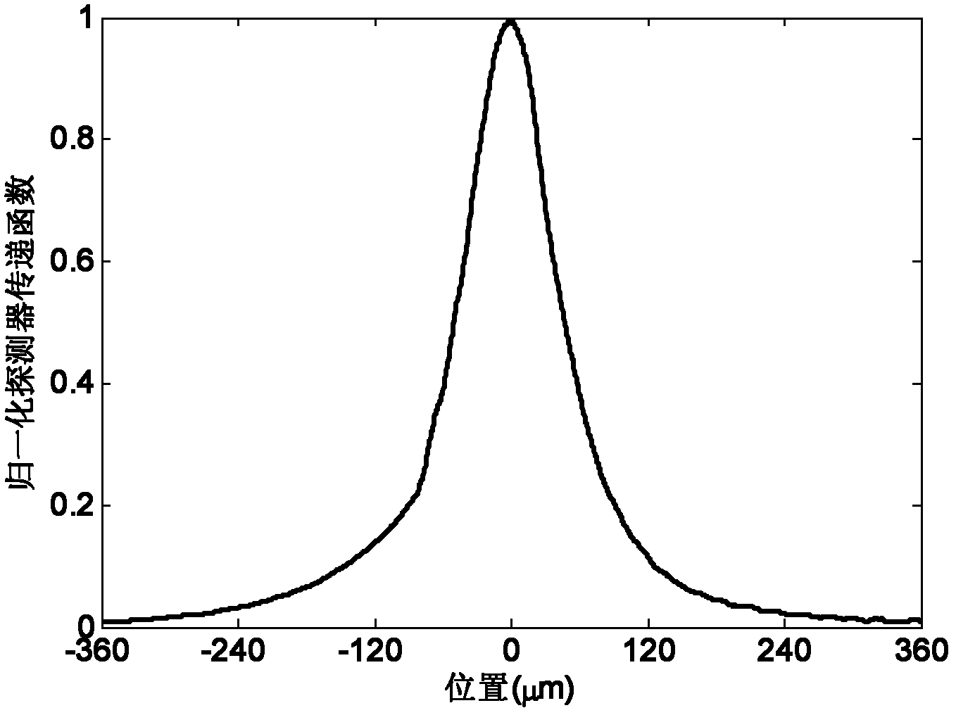
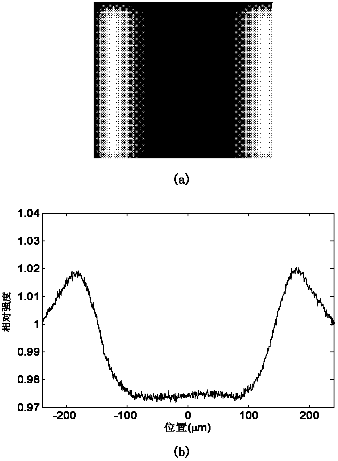
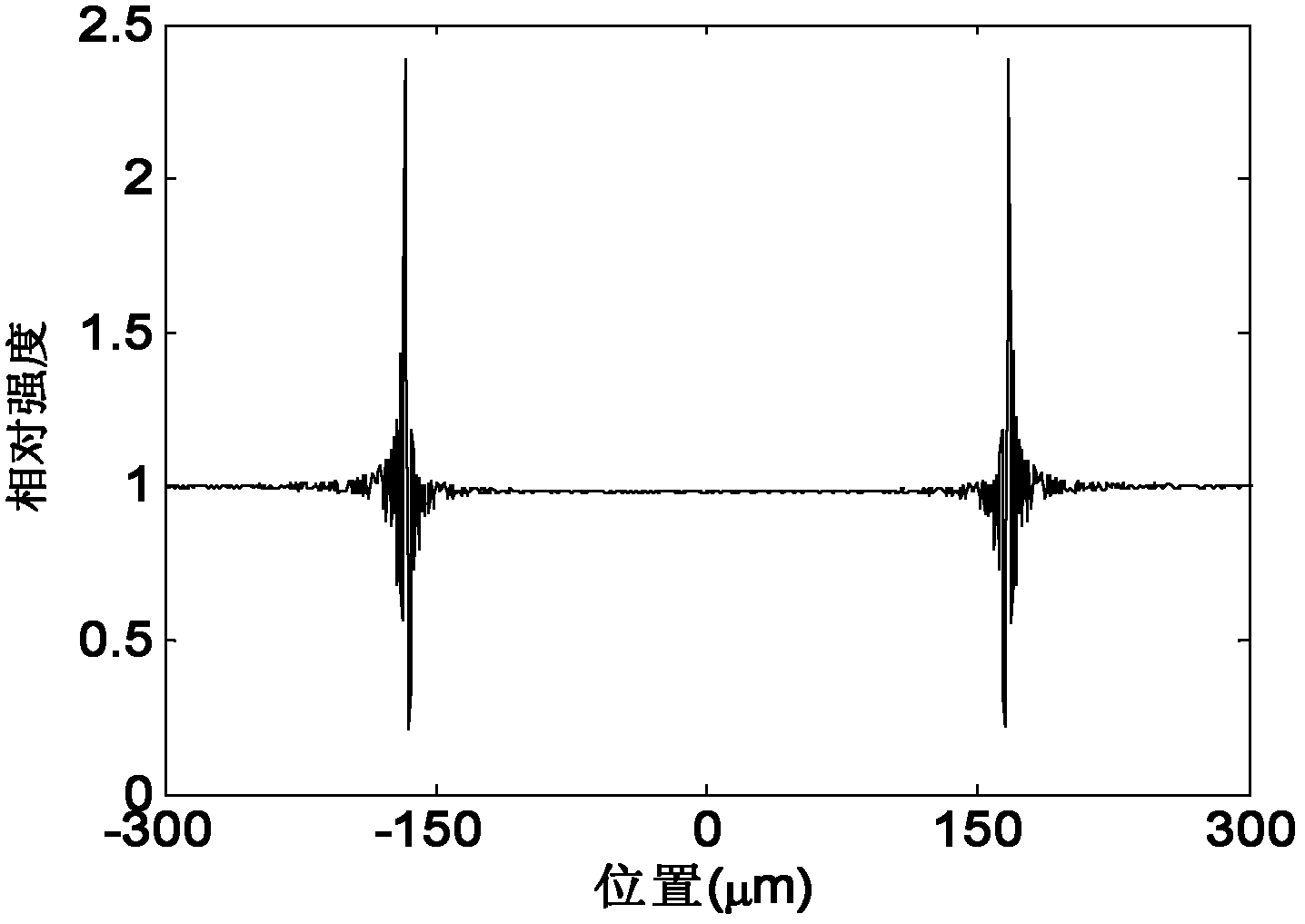
[0032] The experimental imaging system used in the present invention is a Pixarray 100 small animal digital radiography system manufactured by BIOPTICS Corporation of the United States. The detector of the system is a 1024×1024 CCD array with a pixel size of 50 μm×50 μm and 14 gray levels. The horizontal and vertical spatial resolutions are 20 pixels per millimeter. The focal spot size of the X-ray tube is 50 μm. The full width at half maximum of the point spread function of the detector is 110 μm. In the experiment, the working voltage of the X-ray source is 33kVp, and the working current is 0.5mA. The imaging object uses 300μm polyethylene fiber. The two experiments set the distances from the X-ray source to the object as 200cm and 180cm, and the corresponding distances from the object to the detector as 20cm and 40cm. Under the above s...


PUM


Abstract

The invention belongs to the fields of biomedical engineering and medical imaging, relating to an X-ray coaxial phase-contrast imaging method, which comprises the steps of: placing a knife edge device to be appressed onto the surface of a detector, and continuously acquiring n frames of images to obtain a corresponding detector transfer function curve; transforming to obtain a detector transfer function matrix; placing an imaging object and imaging the object; establishing a phase-contrast imaging deterioration model in a matrix manner; selecting a regularization matrix according to the coaxial phase-contrast imaging result of the object and the detector transfer function matrix, and acquiring an optimal regularization parameter corresponding to the regularization matrix by using an L curve method; and calculating a numerical solution of regularization image recovery, of the coaxial phase-contrast imaging result. The X-ray coaxial phase-contrast imaging method disclosed by the invention can be used for effectively increasing the contrast of a phase-contrast image under a deterioration effect and guaranteeing the fidelity of a recovered image.

Description

technical field [0001] The invention belongs to the fields of biomedical engineering and medical imaging, and relates to an X-ray coaxial phase contrast imaging method. Background technique [0002] Breast cancer is currently the number one "killer" of women. According to the statistics of the World Health Organization, in 2000, about 1.2 million women in the world were diagnosed with breast cancer, and 500,000 people died of breast cancer. Breast cancer not only endangers the lives of patients, but also causes damage to female sexual organs, seriously endangering women's physical and mental health. Although China is not a country with a high incidence of breast cancer, its average annual growth rate has been 1-2 percentage points higher than that of high-incidence countries in the past ten years, with an annual growth rate of 3%-4%. Due to the large population in our country, the diagnosis and treatment of breast cancer has become an increasingly serious social problem tha...

Claims


Application Information

Patent Timeline
Patent Type & Authority Applications(China)
IPC IPC(8): A61B6/00
Inventor 周仲兴高峰赵会娟张力新
Owner TIANJIN UNIV
Why Eureka
Try Eureka