A scanning electron microscope sample infection device

A scanning electron microscope and sample technology, which is applied in the preparation of test samples, circuits, discharge tubes, etc., can solve problems such as solution volatilization failure, sample easy to fall off, and affect the accuracy of experimental results, etc., to increase conductivity and secondary electrons Yield, reduced impact on human body and environment, clear and visible sample structure

Active Publication Date: 2017-11-28
GUANGXI MEDICAL UNIVERSITY
View PDF3 Cites 0 Cited by
  • Summary
  • Abstract
  • Description
  • Claims
  • Application Information

AI Technical Summary

Problems solved by technology

In addition, osmic acid can increase the conductivity and secondary electron yield of the sample. The image of the sample fixed by osmic acid has better contrast. It is an excellent fixative widely used in electron microscope technology, but osmic acid is highly toxic and easy to sublimate. Volatile, has a strong stimulating effect on the mucous membranes of the eyes, nose, and throat, and is likely to cause pollution and damage to the human body and the environment
[0004] Conventional scanning electron microscope sample preparation methods have the following problems: 1. It is not suitable for the preparation of some samples with low water content but easy to swell and deform when absorbing water. ;2. Most samples that are not routinely fixed and stained can only be observed at a lower voltage, and the observation and image collection must be rapid, and the quality of the obtained images is poor, which seriously affects the quality of the experimental results and the accuracy of the research; 3. Some scholars take advantage of the easy sublimation and volatilization characteristics of osmic acid, use fine needles or thin threads to insert some samples that are difficult to fix and stain conventionally, and hang them in penicillin bottles and other containers for osmic acid contamination, which can improve the imaging quality of scanning electron microscopy. However, when the performance of the equipment used in the above-mentioned method overcomes the above-mentioned certain defects, it often brings about complex operations, easy to fall off samples, and is not suitable for some brittle and larger samples; 4. Osmic acid has It is highly toxic and easy to sublimate and volatilize. When performing related operations, it must be protected from light and sealed, otherwise it will easily cause the solution to evaporate, pollute the environment, and harm the human body.

Method used


Image

  • A scanning electron microscope sample infection device
  • A scanning electron microscope sample infection device
  • A scanning electron microscope sample infection device

Examples


Embodiment Construction

[0028] The present invention will be described in further detail below in conjunction with the accompanying drawings and specific embodiments.

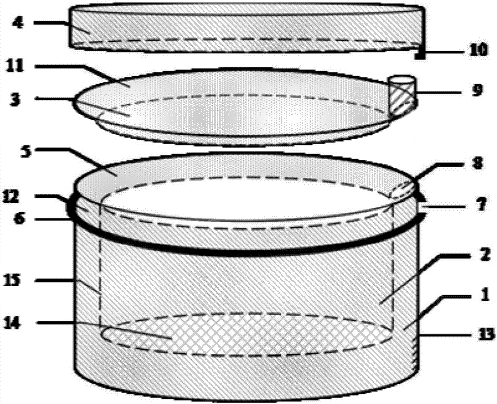
[0029] A scanning electron microscope sample infection device of the present invention such as figure 1 As shown, a scanning electron microscope sample infection device is composed of an outer cylindrical tube 1, a screen set 2, a sealed inner cover 3 and a sealed outer cover 4, forming a closed and cooperative device.

[0030] 1. The structure of each part

[0031] The bottom end of the outer cylindrical tube 1 is closed, and the upper end is open to form a cylindrical lumen, and a positioning ring 6 protruding from the tube wall and a positioning member inlet 7 located on the positioning ring are arranged on the upper middle of the outer wall.

[0032] The sieve set 2 is a cylindrical sieve-like lumen structure with an upper opening of the outer flange 5 of the sieve set, and its lumen is smaller than that of the outer cylindrical ...


PUM

PropertyMeasurementUnit
heightaaaaaaaaaa

Abstract

The invention discloses a scanning electron microscope sample fumigating and dyeing device, which comprises an outer cylindrical tube, a screen sleeve and a seal outer cover, wherein a sample is put into the screen sleeve which is sleeved with an outer cylinder tube cavity in use; and fumigating and dyeing are carried out by covering a seal inner cover, injecting a fumigating and dyeing liquid and covering the seal outer cover. The scanning electron microscope sample fumigating and dyeing device has the beneficial effects that the invention provides the simple, safe and effective scanning electron microscope sample fumigating and dyeing device, so that the defect that an entire structure cannot be observed due to structural damage and loss of a few of samples in conventional preparation processes of scanning electron microscope sample fixing, rinsing, dewatering and the like is overcome; the reagent dosage is saved; the influence of an irritating toxic reagent on a human body and an environment is reduced; the experiment cost and the manpower are reduced; and the experiment success rate and the sample quality are improved.

Description

technical field [0001] The invention relates to a technique for carrying out infection on samples, in particular to an infection device for scanning electron microscope samples. This sample is used for scanning electron microscopy in biomedical research. Background technique [0002] Scanning electron microscope technology is an important means of medical biological research, used to observe the fine structure of the sample surface. For most biological or material samples, traditional SEM specimen preparation methods use chemical or physical methods to fix, dehydrate and dry, and then spray carbon or metal. The specific steps are: use glutaraldehyde fixative to soak the sample for pre-fixation; Rinse the samples after pre-fixation with phosphate buffer; use osmium acid fixative to soak the samples for post-fixation and staining; rinse the samples after post-fixation and staining with phosphate buffer; dehydrate with grade alcohol or acetone; use Use isoamyl acetate or hexa...

Claims


Application Information

Patent Timeline
Patent Type & Authority Patents(China)
IPC IPC(8): H01J37/20G01N1/28
CPCG01N1/28H01J37/20
Inventor 黄远洁胡艳玲
Owner GUANGXI MEDICAL UNIVERSITY
Why Eureka
Try Eureka