Circulating Tumor Cell Protein Typing Kit

A technology of tumor cells and kits, applied in the field of molecular biology, can solve problems such as low sensitivity of flow cytometry, complex operating procedures, false positive results, and insufficient objectivity of non-specificity, so as to improve sensitivity and accuracy, Effect of reducing epithelial cell contamination and increasing fluorescence signal intensity

Active Publication Date: 2019-05-31
SUREXAM BIO TECH
View PDF4 Cites 0 Cited by
  • Summary
  • Abstract
  • Description
  • Claims
  • Application Information

AI Technical Summary

Problems solved by technology

Conventional immunostaining method can only detect one antigen at a time, the operation procedure is complicated and there are false positive results; flow cytometry has low sensitivity and cannot provide cell morphology information; while the CellSearch detection system targets epithelial cell markers dots, CTCs that lost epithelial antigens could not be detected
Fluorescence in situ hybridization is currently used more often, but the accuracy of the test results is affected due to defects such as non-specific antigen-antibody reactions and insufficient objectivity in the judgment of results.

Method used


Image

  • Circulating Tumor Cell Protein Typing Kit
  • Circulating Tumor Cell Protein Typing Kit
  • Circulating Tumor Cell Protein Typing Kit

Examples


Embodiment 1

[0026] There are two types of circulating tumor cell protein typing kits described in this example, those with labeled antibodies and those without labeled antibodies.

[0027] Among them, the circulating tumor cell protein typing kit A with labeled antibodies mainly includes:

[0028] 1. Capture antibody

[0029] The capture antibody is connected to the marker protein and the marker antibody, and can specifically bind to the marker protein and the marker antibody. The capture antibody is a monoclonal antibody with high purity, and the selected capture antibodies do not interfere with each other in their reaction characteristics.

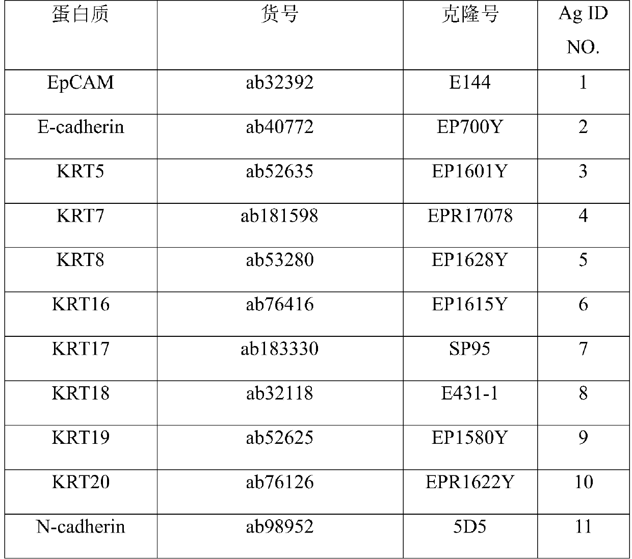
[0030]In this embodiment, the marker proteins are divided into three cell types, which are epithelial cell marker proteins, mesenchymal cell marker proteins and leukocyte marker proteins, and the epithelial cell marker proteins are: EPCAM, E-cadherin, KRT5, KRT7 , KRT8, KRT16, KRT17, KRT18, KRT19, KRT20; the interstitial cell marker proteins are: N...

Embodiment 2

[0050] Example 2 Using the kit A in Example 1 to detect circulating tumor cells in human peripheral blood

[0051] The formula of described various solutions is as follows:

[0052]

[0053]

[0054] All the antibodies in the list of corresponding marker proteins in Example 1 were used in the capture mixed solution and the chromogenic mixed solution in this example.

[0055] 1. Sample pretreatment, filter CTCs onto the filter membrane

[0056] 1. Preserve the blood sample in the sample preservation tube with preservation solution, centrifuge at 600×g for 5 minutes, discard the supernatant, and remove the red blood cells.

[0057] 2. Add 200uL magnetic bead antibody, 4mL PBS and 1mL fixative, vortex and mix well, and let stand at room temperature for 8min.

[0058] 3. Sample filtration: Transfer the liquid in the sample storage tube to the filter, turn on the vacuum filtration pump to pump out the liquid; add 4mL PBS to the storage tube, wash the tube wall and filter th...

Embodiment 3

[0109] Example 3: Application of labeled antibodies

[0110] 1. Design of kit preparation (signal detection components)

[0111] There are two options for the signal detection component of the kit of the present invention, 1) the capture antibody combined with the marker protein is directly modified with a fluorescent group; 2) the marker protein is combined with the capture antibody and then combined with the labeled antibody, and the labeled antibody is modified with a fluorescent group group. These two signal detection components can both realize signal amplification and detect normal signals. Among them, the detection signal is more stable and the effect is better when the labeled antibody modified by the fluorescent group is used.

[0112] Taking the two signal detection components of the kit for detecting epithelial cell marker proteins as an example, the specific design is shown in Table 7. That is, the kit consists of:

[0113] Experimental group 1: the capture ant...


PUM


Abstract

The invention relates to a protein typing kit of circulating tumor cells. The kit comprises capture antibodies against different CTCs marker proteins and labeled antibodies against different CTCs marker proteins. The CTCs marker proteins comprise epithelial cell marker proteins and interstitial cell marker proteins. The epithelial cell marker proteins are selected from EPCAM, E-cadherin, KRT5, KRT7, KRT-8, KRT-16, KRT-17, KRT-18, KRT-19, KRT-20. The interstitial cell marker proteins are selected from N-cadherin, vimentin, fibronectin, MMP2, MMP3, MMP9, AKT2, ZEB1, FOXC1, FOXC2, SNAI1, SNAI2, SERPINE1. The marker proteins selected in the invention can realize detection of single marker protein, and a single marker protein can be used with other marker proteins for the detection. Therefore, circulating tumor cells can be fully detected, and the cell type can be distinguished. And then, false negative results caused by difference of expression levels of some marker proteins among individual circulating tumor cells are avoided.

Description

technical field [0001] The invention belongs to the field of molecular biology, relates to medicine and biotechnology, in particular to a circulating tumor cell protein typing kit. Background technique [0002] The ability of tumor cells to acquire invasion and distant metastasis is the main reason for the death of patients. Circulating tumor cells (CTCs) were first discovered in 1869. They originate from primary tumors or metastatic tumors. They acquire the ability to escape from the basement membrane and enter blood vessels by invading the tissue matrix. Generic term for tumor cells. The current theory of tumor metastasis believes that tumor cells need to break away from the primary tumor, break through the vascular barrier and enter the blood or lymphatic circulation to form metastases in the distance, so the formation of CTCs may be the early stage of tumor metastasis. CTC is a group of heterogeneous cells. According to the expression of marker proteins, it is mainly d...

Claims


Application Information

Patent Timeline
Patent Type & Authority Patents(China)
IPC IPC(8): G01N33/68
CPCG01N33/56966G01N33/68
Inventor 刘苏燕吴诗扬
Owner SUREXAM BIO TECH
Why Eureka
Try Eureka