OCT (Optical Coherence Tomography) image segmentation method based on random forest and composite active curve

A random forest and image layer technology, applied in the field of medical image processing algorithms, can solve problems such as leakage to the neighborhood, low contrast of the image retina layer, layer segmentation failure, etc.

Active Publication Date: 2017-11-24
SUZHOU UNIV
View PDF3 Cites 5 Cited by
  • Summary
  • Abstract
  • Description
  • Claims
  • Application Information

AI Technical Summary

Problems solved by technology

This results in low contrast and blurred boundaries between retinal layers in OCT images, and also highly variable retinal layer structure
Therefore, layer segmentation may fail using traditional surface detection methods such as image search algorithms, and meanwhile, effusion segmentation using traditional methods such as region growing may also easily leak into the neighborhood

Method used


Image

  • OCT (Optical Coherence Tomography) image segmentation method based on random forest and composite active curve
  • OCT (Optical Coherence Tomography) image segmentation method based on random forest and composite active curve
  • OCT (Optical Coherence Tomography) image segmentation method based on random forest and composite active curve

Examples


Embodiment 1

[0114] The OCT image layer segmentation method based on random forest and compound activity curve in this embodiment includes:

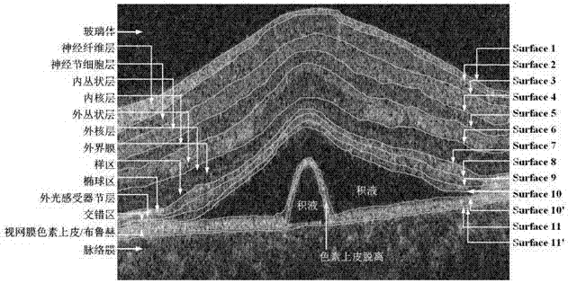
[0115] like figure 1 As shown, training the OCT image features to train the random forest classifier, when training the random forest classifier, the central serous retinopathy retinal OCT image is divided into 8 regions; region 1: nerve fiber layer; region 2: ganglion cell layer Area 3: inner plexiform layer; area 4: inner core layer; area 5: outer plexiform layer; area 6: outer nuclear layer + outer membrane + sample area; area 7: ellipsoid area + outer photoreceptor node layer + interlaced Area + retinal pigment epithelium / Bruch; area 8 (class 8): vitreous + choroid; the upper surface layer of area 1 Surface1, the upper surface of area 2 Surface2... The upper surface of area 8, Surface1 is referred to as SF1, the same is Surface2 SF2 for short...Surface8 is abbreviated as SF8. For details, see figure 1 shown.

[0116] The specific methods of ...

Embodiment 2

[0125] This embodiment is based on the OCT image layer segmentation method of random forest and compound activity curve. On the basis of Embodiment 1, the obtained OCT image feature training random forest classifier specifically includes:

[0126] The central serous retinopathy retinal OCT image was segmented into 8 regions; region 1: nerve fiber layer; region 2: ganglion cell layer; region 3: inner plexiform layer; region 4: inner nuclear layer; region 5: outer plexiform layer layer; area 6: outer nuclear layer + outer membrane + sample area; area 7: ellipsoid area + outer photoreceptor nodal layer + interlaced area + retinal pigment epithelium / Bruch; area 8 (class8): vitreous + choroid; area The upper surface layer of 1, Surface1, the upper surface of area 2, Surface2... The upper surface of area 8, Surface1 is referred to as SF1, and similarly Surface2 is referred to as SF2...Surface8 is referred to as SF8 for details. figure 1 The specific division shown is not limited t...


PUM


Abstract

The invention relates to an OCT (Optical Coherence Tomography) image segmentation method based on a random forest and a composite active curve, and is designed for accurately segmenting a retina layer and hydrops. The OCT image segmentation method based on the random forest and the composite active curve comprises the following steps that: obtaining OCT image features to train a random forest classifier, and obtaining a final SF1 through a composite active curve algorithm; extracting 24 features of the OCT image; and using the random forest classifier. The OCT image segmentation method based on the random forest and the composite active curve has the advantages of being simple in operation and accurate in detection results. The existing problems of low identification rate, poor segmentation effect and the like of a lesion OCT image segmentation algorithm are overcome.

Description

technical field [0001] The invention belongs to the field of medical image processing algorithms, in particular to an OCT image layer segmentation method based on random forest and compound activity curve. Background technique [0002] Central serous retinopathy is a severe and complex retinal disease that can easily lead to blindness. Central serous retinopathy occurs mainly as the accumulation of subretinal serous fluid in the staggered area, which may also lead to retinal pigment epithelial detachment. In addition to serous accumulation, retinal pigment epithelium detachment may occur under the serous or near the center of the macula. This fluid causes swelling of the retinal layers, which may suddenly change in thickness and optical intensity. Thus, central serous retinopathy is a common disease of the macula, which is responsible for central vision. Therefore, quantitative analysis of central serous retinopathy is of great significance in retinal research. [0003] ...

Claims


Application Information

Patent Timeline
Patent Type & Authority Applications(China)
IPC IPC(8): G06T7/11G06K9/62
CPCG06T7/11G06T2207/30096G06T2207/30041G06F18/24323
Inventor 向德辉
Owner SUZHOU UNIV
Why Eureka
Try Eureka