A medical image processing system and method

A medical image and processing system technology, applied in the field of medical image processing systems, can solve the problems of low accuracy and slow speed of lesion detection methods, and achieve the effects of ensuring accuracy, improving detection speed, and improving acquisition speed

Active Publication Date: 2021-12-10
SHANGHAI UNITED IMAGING HEALTHCARE
View PDF9 Cites 0 Cited by
  • Summary
  • Abstract
  • Description
  • Claims
  • Application Information

AI Technical Summary

Problems solved by technology

[0004] The embodiment of the present invention provides a medical image processing system and method to solve the problems of low accuracy and slow speed of the existing lesion detection method

Method used


Image

  • A medical image processing system and method
  • A medical image processing system and method
  • A medical image processing system and method

Examples


Embodiment 1

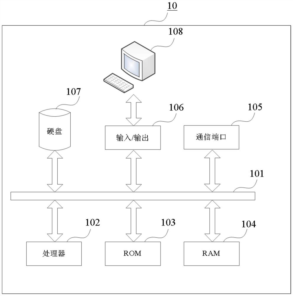
[0054]Embodiment 1 of the present invention proposes a medical image processing system, which can be used to implement the specific methods disclosed in some embodiments of the present invention. The specific system in this embodiment uses a functional block diagram to show a hardware platform including a display module. In some embodiments, the medical image processing system can realize the specific implementation of some embodiments of the present invention through its hardware device, software program, firmware and their combination. In some embodiments, the medical image processing system may be a general purpose computer, or a medical imaging device with image processing functions. The medical imaging equipment may be electrocardiography (electrocardiography), digital radiography (digitalradiography, DR) equipment, magnetic resonance imaging (magnetic resonance imaging, MRI) equipment, computer tomography (computed tomography, CT) equipment, positron emission type comput...

Embodiment 2

[0079] Figure 5It is a flow chart of a medical image processing method provided by Embodiment 2 of the present invention, and the instructions involved in the method can also be executed by the processor 102 . The technical solution of this embodiment can be applied to detect lesions in medical images, and further screen lesions to remove false positives. The method specifically includes the following operations:

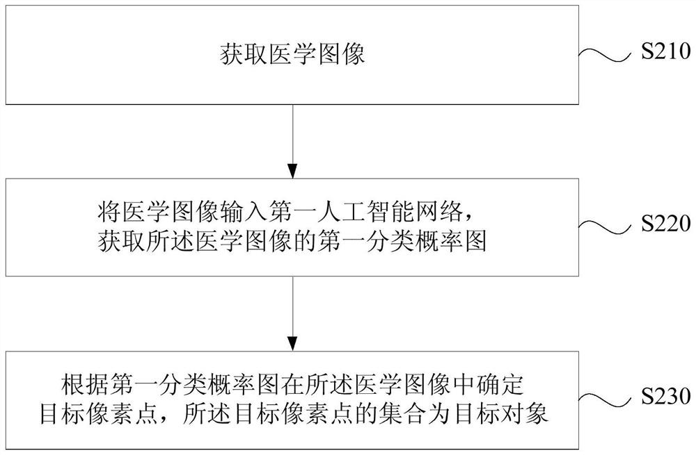
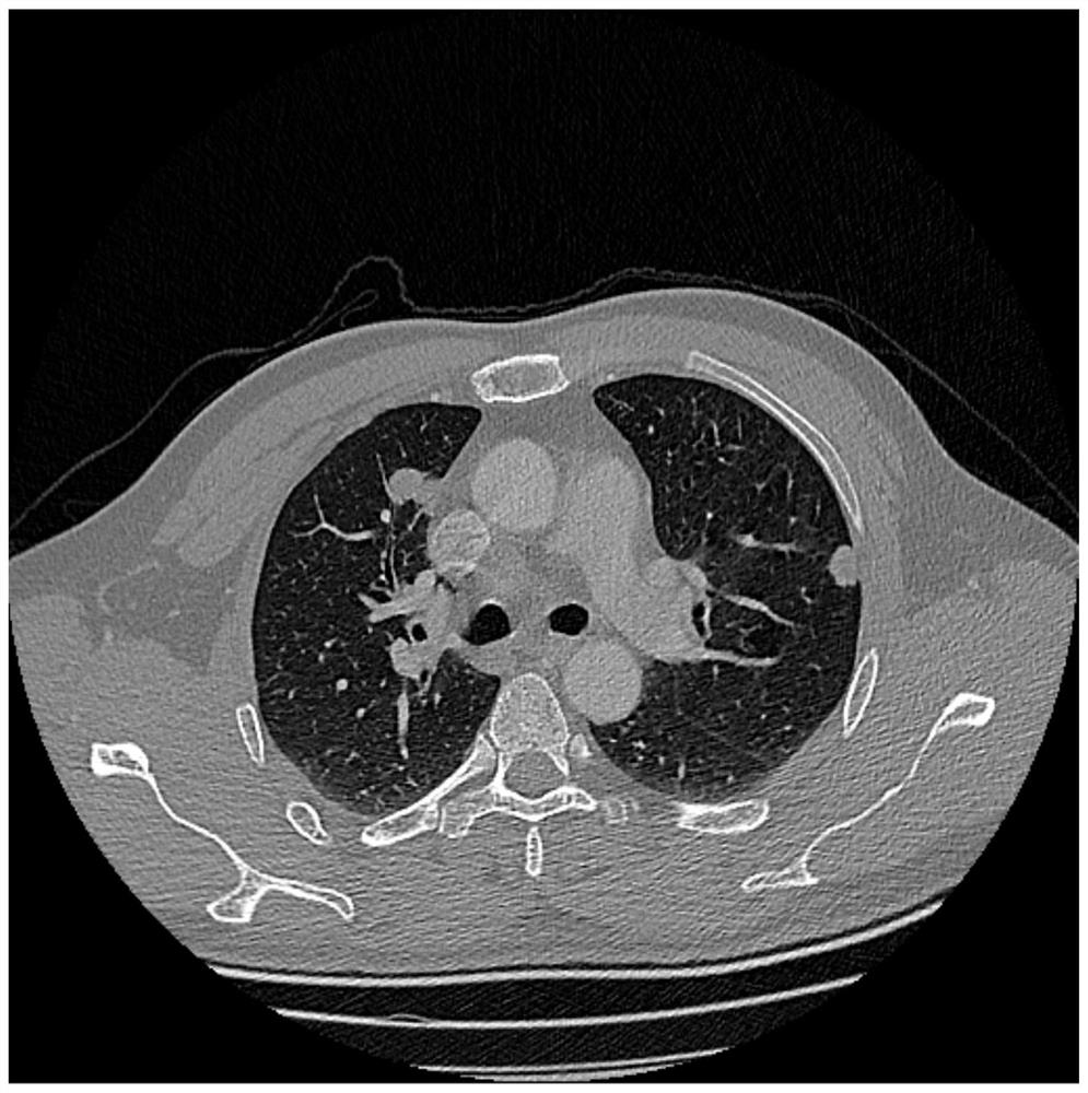
[0080] S510. Acquire a medical image, where the medical image may include multiple pixels. In this embodiment, the medical image is a CT lung image, and an initial segmentation is performed on the medical image to obtain lung regions.

[0081] S520. Input the medical image into the first artificial intelligence network, and obtain a first classification probability map of each pixel of the medical image.

[0082] S530. Determine target pixel points in the medical image according to the first classification probability map.

[0083] S540. Remove false positive p...

Embodiment 3

[0100] Figure 7 It is a flowchart of a medical image processing method provided by Embodiment 3 of the present invention. The technical solution of this embodiment is further optimized on the basis of any of the foregoing embodiments. The method of this embodiment includes:

[0101] S710. Acquire a medical image, where the medical image includes multiple pixels.

[0102] S720. Input the medical image into a first artificial intelligence network to classify each pixel of the medical image into target pixels and non-target pixels. The first artificial intelligence network is based on medical image samples and corresponding Obtained by training the target pixels.

[0103] In this embodiment, the first artificial intelligence network selects a region proposal network (region proposal network, RPN) to input the medical image into the first artificial intelligence network, so as to classify each pixel of the medical image into target pixels and non-target pixels. The target pix...


PUM


Abstract

The embodiment of the invention discloses a medical image processing system and method. The system includes: one or more processors; memory for storing one or more programs, when the one or more programs are executed by the one or more processors, the one or more processors Realization: obtain medical images; input the medical images into the first artificial intelligence network, and obtain the first classification probability map of the medical images, and each point in the first classification probability map corresponds to the pixel points of the medical images Correspondingly, the first artificial intelligence network is pre-trained based on medical image samples and corresponding target pixels; the target pixels are determined in the medical image according to the first classification probability map, and the target pixels are Collection is the target object. The embodiments of the present invention improve the detection rate of target pixels and ensure the accuracy of target object detection.

Description

technical field [0001] Embodiments of the present invention relate to medical image processing technologies, and in particular to a medical image processing system and method. Background technique [0002] When performing medical image detection, there are usually two situations in the detection results, one is the real target area, that is, the body position of the actual lesion; the other is a false positive, that is, the body position that does not actually have a lesion but the detection result is a lesion. Therefore, when performing medical image detection, filtering out false positives is crucial for correctly detecting the location of lesions. [0003] Existing methods for lesion detection first use a sliding window for initial detection, and then use a convolutional neural network to remove false positives. Due to the fact that the proportion of lesions and false positives in the image is very small, when the size of the window is too large, there will be problems o...

Claims


Application Information

Patent Timeline
Patent Type & Authority Patents(China)
IPC IPC(8): G06T7/00G06K9/62
CPCG06T7/0012G06T2207/30061G06T2207/20084G06T2207/10081G06T2207/20032G06F18/241
Inventor 王季勇李强
Owner SHANGHAI UNITED IMAGING HEALTHCARE
Why Eureka
Try Eureka