
Analysis method of myocardial shapes, motions and deformations in two-dimensional echocardiogram sequence

A technology of echocardiography and myocardial movement, applied in ultrasound/sound wave/infrasonic wave diagnosis, sound wave diagnosis, infrasonic wave diagnosis, etc., can solve unresolved myocardial movement and deformation information, difficult to meet the requirements of real-time processing, and improve clinical diagnosis function Limited and other issues

Active Publication Date: 2018-05-18
ZHEJIANG CHINESE MEDICAL UNIVERSITY
View PDF6 Cites 3 Cited by
  • Summary
  • Abstract
  • Description
  • Claims
  • Application Information

AI Technical Summary

Problems solved by technology

The shortcomings of this method are: (1) PET heart image is a three-dimensional image, and the amount of data that needs to be processed at each moment in the heart cycle is very huge, which is difficult to meet the requirements of clinical practice for real-time processing
(2) Only the heart contour is extracted by using PET heart images, but the myocardial motion and deformation information is not analyzed, which has limited effect on improving clinical diagnosis

Method used


Image

  • Analysis method of myocardial shapes, motions and deformations in two-dimensional echocardiogram sequence
  • Analysis method of myocardial shapes, motions and deformations in two-dimensional echocardiogram sequence
  • Analysis method of myocardial shapes, motions and deformations in two-dimensional echocardiogram sequence

Examples


Embodiment Construction

[0085] The present invention will be further described below in conjunction with specific examples, but the protection scope of the present invention is not limited thereto.

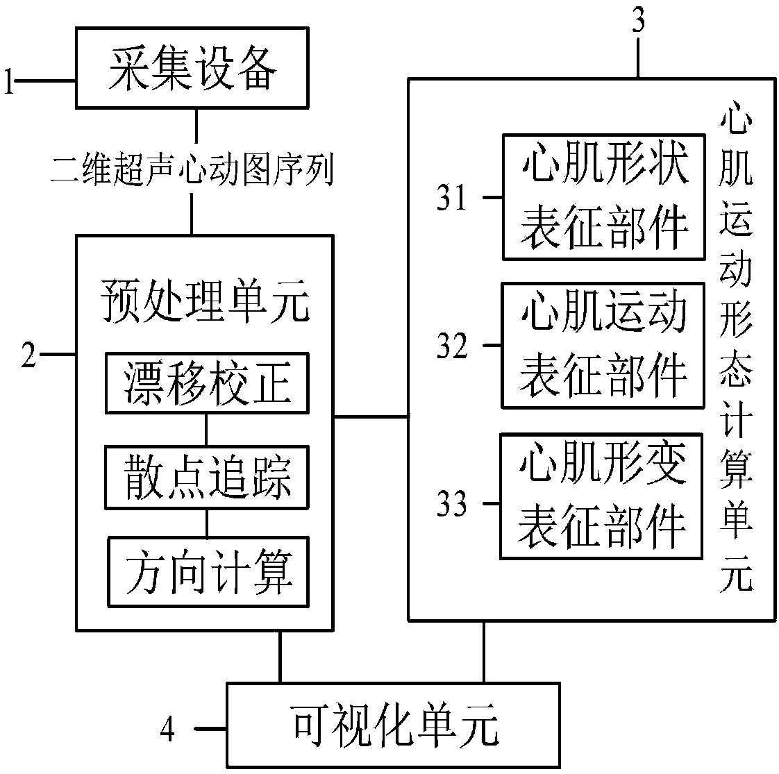
[0086] Example 1. Myocardial shape, motion and deformation analysis system in two-dimensional echocardiography sequence, such as figure 1 As shown, it includes an acquisition device 1 , a preprocessing unit 2 , a myocardial motion shape calculation unit 3 and a visualization unit 4 connected in sequence, wherein the preprocessing unit 2 is connected to the visualization unit 4 .

[0087] The acquisition device 1 is used for acquiring a two-dimensional echocardiogram.

[0088] The preprocessing unit 2 is used to import the sequence data of the two-dimensional echocardiogram in the acquisition device 1 and preprocess the sequence data; in this embodiment, the preprocessing unit 2 outputs each point of the cardiac cycle myocardial wall (that is, the myocardial point ) coordinates to the myocardial motion s...


PUM


Abstract

The invention discloses an analysis method of myocardial shapes, motions and deformations in a two-dimensional echocardiogram sequence, including the following steps: first, an analysis two-dimensional echocardiogram sequence to be analyzed is preprocessed by a preprocessing unit, so that the coordinates of each point of a myocardial wall in a cardiac cycle are obtained, and the direction of the myocardial wall at each time point of the myocardial wall in the cardiac cycle is displayed on a visualization unit; then, a myocardial motion pattern calculation unit calculates myocardial shapes, displacements, velocities and longitudinal strains according to the coordinates of each point of the myocardial wall in the cardiac cycle, so that the cardiac mechanics is intuitively understood, particularly the myocardial shapes, motions and deformations. The analysis method of the myocardial shapes, motions and deformations in the two-dimensional echocardiogram sequence analyzes images at each time point in the cardiac cycle, thereby displaying the myocardial shapes, motions and deformations in a cardiac motor process, satisfying requirements of clinical practice for a real time processing, being capable of effectively avoiding accidental risks of some static images in clinic and convenient for clinicians to make right judgments.

Description

technical field [0001] The invention relates to the fields of medical image processing and intelligent health management, in particular to a method for analyzing myocardial shape, movement and deformation in a two-dimensional echocardiogram sequence. Background technique [0002] Cardiac diseases such as coronary heart disease are currently one of the main causes of human death, and medical imaging technology plays an important role in the diagnosis and treatment of heart diseases. Echocardiography is noninvasive, safe and reliable, and is widely used in clinical diagnosis of heart disease. The study of the motion of the heart by 2D echocardiography, especially the study of the left ventricle, is at the center of attention. Although the motion of the heart can be observed directly from two-dimensional real-time ultrasound images, there is still potential for major improvements in its diagnostic quality. Since real-time observation is basically a qualitative evaluation, it ...

Claims


Application Information

Patent Timeline
IPC IPC(8): A61B8/00
CPCA61B8/0883A61B8/5207A61B8/5223
Inventor 赖小波徐小媚方纯洁方颖吕莉莉
Owner ZHEJIANG CHINESE MEDICAL UNIVERSITY
Learn More