
Application of FOXM1 inhibitor FDI-6 in resistance of hepatic fibrosis
A FDI-6, anti-hepatic fibrosis technology, applied in the digestive system, medical preparations containing active ingredients, organic active ingredients, etc.
- Summary
- Abstract
- Description
- Claims
- Application Information
AI Technical Summary
Problems solved by technology
Method used
Image

Examples
Embodiment 1
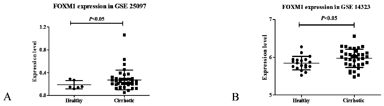
[0035] Example 1: Analyzing clinical samples through the more severe liver fibrosis—cirrhosis tissue gene expression profile (gene expression omnibus, GEO) database on the Internet.
[0036] Experimental materials: two liver cirrhosis databases, GSE25097 and GSE 14323.
[0037] Experimental method: The GEO database GSE25097 and GSE 14323 of liver cirrhosis were investigated online to screen the differential expression of FOXM1.
[0038] Experimental results: Both databases showed that the expression of FOXM1 in cirrhotic tissues was significantly higher than that in normal tissues.
Embodiment 2
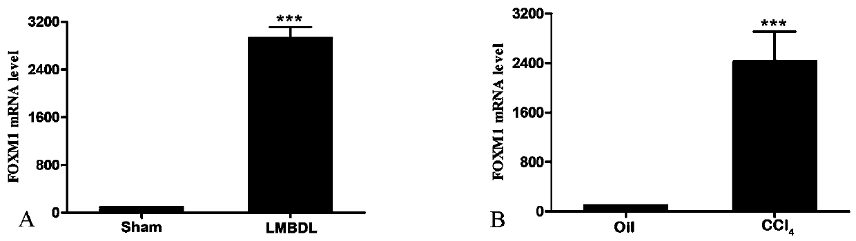
[0039] Example 2: Left middle common bile duct ligation (LMBDL) induced liver fibrosis in mice.
[0040] Experimental materials: C57L / 6 mice, 3% sodium pentobarbital solution, surgical instruments.
[0041] Experimental method: 12-week-old male C57BL / 6 mice were anesthetized by intraperitoneal injection of 3% pentobarbital sodium solution, the skin and muscle layer of the abdominal cavity were incised, the common bile duct was freed, the left middle common bile duct was ligated, and the abdominal cavity was sutured.
[0042] Experimental results: After about 10 days, ALT and AST were significantly increased, and HE and Sirius red staining showed obvious liver fibrosis.
Embodiment 3
[0043] Embodiment 3: carbon tetrachloride (CCl 4 ) induced liver fibrosis in mice.
[0044] Experimental materials: C57L / 6 mice, CCl 4 solution, olive oil, 1ml syringe.
[0045] Experimental method: 12-week-old male C57BL / 6 mice were intraperitoneally injected with CCl diluted in olive oil 4 Solution (by volume ratio, CCl 4 : olive oil = 1: 3) twice a week.
[0046] Experimental results: After about 4 weeks, both ALT and AST were significantly increased, and HE and Sirius red staining showed obvious liver fibrosis.
PUM
 Abstract
Description
Claims
Application Information