Method and system for supporting medical personnel during a resection, and computer program product

A technology for medical staff and data sets, applied in computer-aided surgery, computer-aided planning/modeling, surgical navigation systems, etc., can solve problems such as expensive, patient burden, and time-consuming

Pending Publication Date: 2021-03-19
SIEMENS HEALTHCARE GMBH
View PDF0 Cites 0 Cited by
  • Summary
  • Abstract
  • Description
  • Claims
  • Application Information

AI Technical Summary

Problems solved by technology

Both are very expensive and time-consuming, and may impose an additional burden on the respective patients
Furthermore, this imaging method cannot usually be used without interrupting the corresponding intervention, i.e. it can only be applied step by step, respectively

Method used


Image

  • Method and system for supporting medical personnel during a resection, and computer program product
  • Method and system for supporting medical personnel during a resection, and computer program product
  • Method and system for supporting medical personnel during a resection, and computer program product

Examples


Embodiment Construction

[0061] The examples described below are preferred embodiments of the present invention. In this exemplary embodiment, the components described in the embodiments each represent individual features of the invention which are to be considered independently of each other, which also respectively develop the invention independently of each other, and are therefore also to be considered individually or together with the illustrated Combinations of various are considered to form part of the present invention. Furthermore, the described embodiments can also be supplemented by further features of the invention which have already been described.

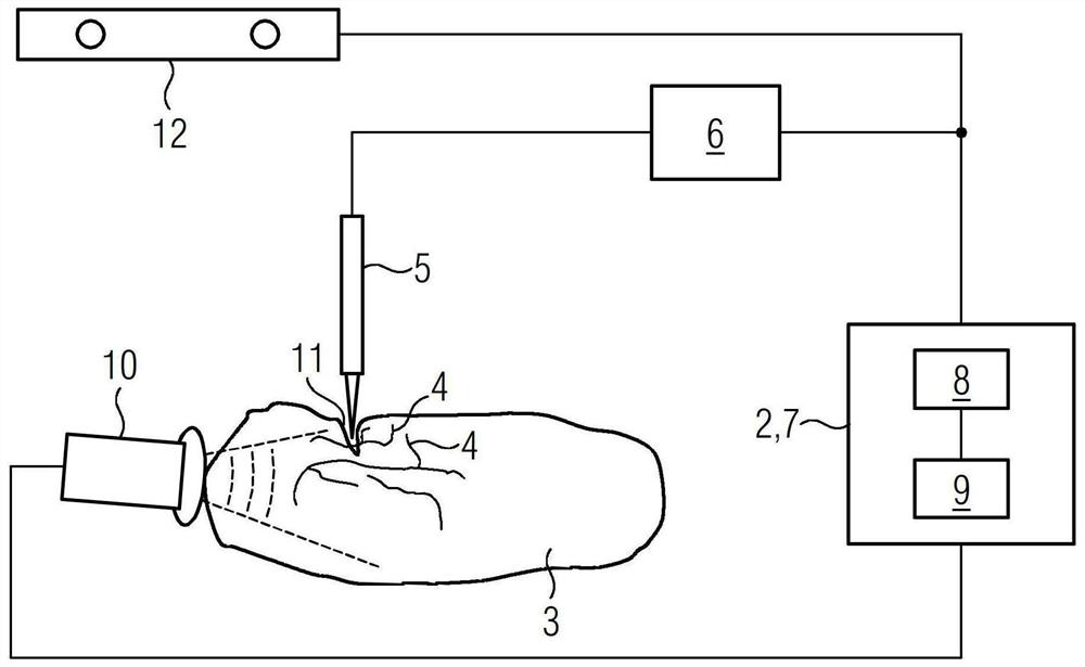
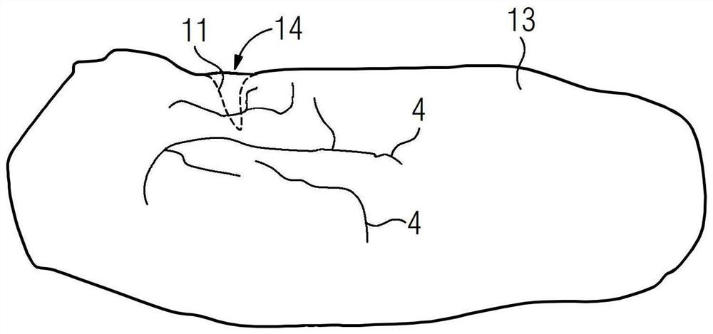
[0062] In the figures, elements that are identical, have the same function or correspond to one another are identified with the same reference numerals.

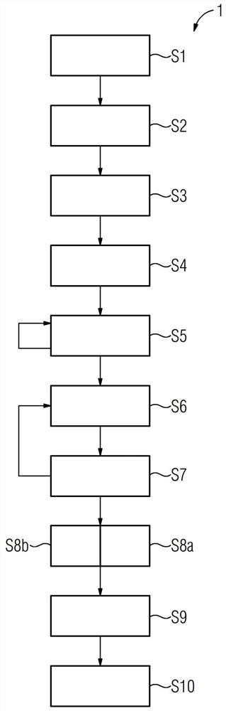
[0063] figure 1 Exemplary illustration of a schematic procedure for a method for supporting medical personnel during resection figure 1 . Here, the method steps S1 to S10 that are carri...


PUM


Abstract

The invention relates to a method (1) for supporting medical personnel during a resection. The invention also relates to a corresponding system (2) and a corresponding computer program product (1, 9).In the method, a preoperative three-dimensional (3D) data set (13) of an examination object (3) on which the resection is to be carried out is acquired. Furthermore, a resection surface (11) is acquired by ultrasound to generate an ultrasound data set describing the resection surface (11). The ultrasound data set is registered with the respective current 3D data set (13). The current 3D data set(13) is updated with the resection surface (11) acquired by ultrasound.

Description

technical field [0001] The invention relates to a method for supporting medical staff during resection, a corresponding computer program product and a system for carrying out such a method. Background technique [0002] Preoperative imaging has established itself as an essential part of device-supported and computer-supported medicine. The preoperatively acquired image data can be used, for example, as a basis for preoperative planning and for intraoperative navigation. However, once the resection has started within the scope of the corresponding operation or intervention (for example, a liver tumor), the preoperative image data or the resulting 3D model (for example of the liver) are no longer current, since they do not represent the actual Performed resection. [0003] Previous methods for estimating intraoperative cutting planes have used optical stereo reconstruction in which stereo camera images of the cutting planes are recorded and from which an attempt has been mad...

Claims


Application Information

Patent Timeline
Patent Type & Authority Applications(China)
IPC IPC(8): A61B34/10A61B34/20A61B90/00
CPCA61B34/10A61B34/20A61B90/37A61B2034/105A61B2034/2063A61B2034/2055A61B2090/374A61B2090/378A61B2090/3762A61B2034/107A61B2034/102
Inventor A.阿尔扎加A.雷根斯伯格S.瓦尔特M.维茨
Owner SIEMENS HEALTHCARE GMBH
Why Eureka
Try Eureka