Method and apparatus for acquiring and displaying a medical instrument introduced into a cavity organ of a patient to be examined or treated

a medical instrument and cavity organ technology, applied in the field of medical instruments acquisition and display, can solve the problems that the anatomy of the patient can only be imaged very inadequately in the fluoroscopic image during the intervention, and achieve the effect of facilitating the recognition of the position of the instrumen

Active Publication Date: 2005-08-02
SIEMENS HEALTHCARE GMBH
View PDF14 Cites 125 Cited by
  • Summary
  • Abstract
  • Description
  • Claims
  • Application Information

AI Technical Summary

Benefits of technology

[0013]An object of the present invention is to provide a method and an examination or treatment device that facilitates the recognition of the position of the instrument.
[0015]Due to the intracorporeal acquisition of the 2D ultrasound images and the generation of the 3D ultrasound image dataset based thereon, the method and device enable the generation of a three-dimensional inside view of the cavity organ, substantially during the intervention, so the instrument can already be introduced into the cavity during the ultrasound image acquisition or shortly thereafter. When a 3D ultrasound image dataset set has been generated, then the acquisition of the instrument introduced into the cavity organ and the determination of corresponding coordinates follows, the latter, due to their registration with the coordinate system of the 3D ultrasound image dataset, enabling a presentation of the instrument in the 3D reconstruction image at the monitor that is exact as to the instrument's position and orientation. The invention advantageously enables the instrument guidance, i.e., for example, the guidance of a catheter advanced into the heart, to be supported by high-resolution 3D imaging, with the 3D image data being intraoperatively generated, so that the current anatomical conditions are reproduced in the three-dimensional image. Ultrasound is employed as the imaging modality, this enabling the generation of an adequately high-resolution 3D ultrasound image dataset with no radiation load on the patient due to the ultrasound image acquisition. On the basis of the registered instrument acquisition, the positionally exact mixing of the instrument, i.e., for example, the catheter tip or a catheter tip section of a suitably long dimension into the three-dimensional presentation can ensue. Overall, the physician obtains an organ exposure produced quasi in situ with the displayed instrument, which enables a significantly better orientation for the physician in the framework of the subsequent navigation.
[0018]If the examination region is a rhythmically or arrhythmically moving region, for example the heart, then for an exact presentation only those 2D ultrasound images that show the cavity organ in the respectively same motion phase can be employed for generating the 3D ultrasound image dataset, and thus the 3D reconstruction image. In order to enable this, in such an instance that an indication of the motion phase of the cavity organ is acquired for the 2D ultrasound images, and only those 2D ultrasound images that are acquired in the same motion phase are employed for generating the 3D ultrasound image dataset. This makes it possible to reconstruct the volume image on the basis of isophase images. It is expedient when—in addition to the motion phase—the point in time of the acquisition of the 2D ultrasound images also is acquired, and only those 2D ultrasound images that are also acquired at the same point in time are employed for generating the 3D ultrasound image dataset.
[0022]In a further embodiment of the invention, during the acquisition of the 2D ultrasound images, a 3D reconstruction image generated on the basis of the essentially isochronically generated 3D ultrasound image dataset is output at the monitor and shows the position of the ultrasound fan output by the ultrasound generating device or the position and orientation of the ultrasound generating device in real time. This mixing makes it possible for the cardiologist to modify the position and orientation of the ultrasound acquisition catheter such that the subsequently acquired 2D ultrasound images contain medically relevant anatomical regions (for example, the target spot of a following ablation and the like). Moreover, the cardiologist thus can perceive the direction in which the catheter-like acquisition device must be navigated in order to complete or, respectively, designationally expand the 2D ultrasound image dataset.

Problems solved by technology

A problem associated with such a procedure from a medical / technical point of view is that although the catheter can be visualized very exactly and highly resolved during the X-ray supervision in one or more fluoroscopic images—also called fluoro images—during the intervention, the anatomy of the patient can be imaged only very inadequately in the fluoroscopic images during the intervention.

Method used


Image

  • Method and apparatus for acquiring and displaying a medical instrument introduced into a cavity organ of a patient to be examined or treated
  • Method and apparatus for acquiring and displaying a medical instrument introduced into a cavity organ of a patient to be examined or treated

Examples


Embodiment Construction

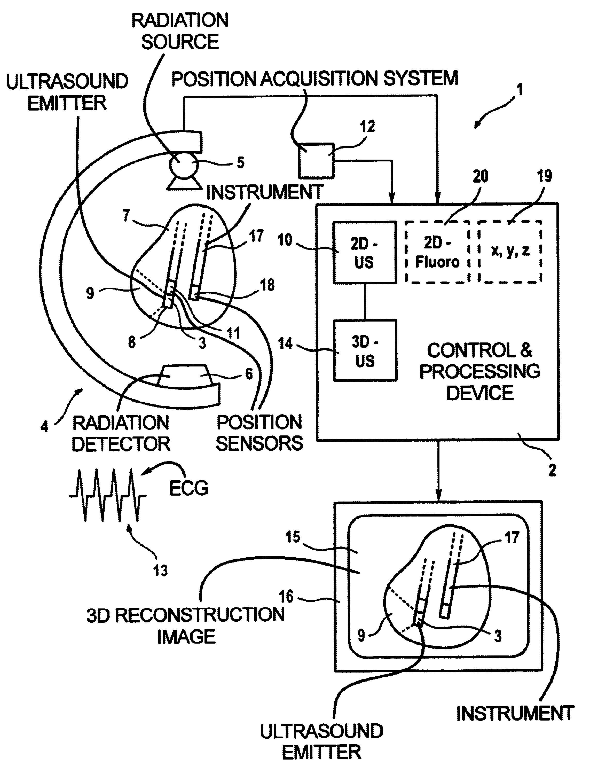
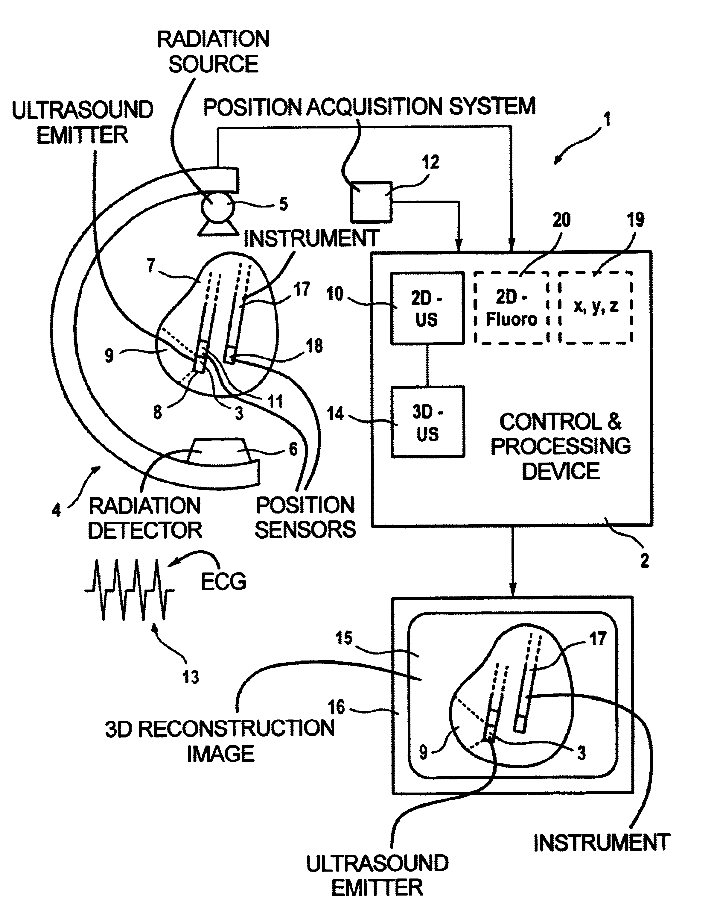
[0024]In the form of a schematic illustration, the drawing shows an inventive medical examination and / or treatment apparatus 1. In the illustrated example, this includes a central control and processing device 2, an ultrasound acquisition device 3 that is fashioned as a catheter-like applicator and that is in communication with the control and processing device 2, as well as a radiation image acquisition device 4 having a radiation source 5 and a radiation detector 6 with which, for example, 2D fluoroscopic images of an examination region in the form of a cavity organ 7 are possible—a heart (shown enlarged) in this case.

[0025]2D ultrasound images 10 of the inside of the cavity organ 7 are acquired with the ultrasound acquisition device 3, which has an ultrasound transmitter 8 that emits an ultrasound fan 9. As can be seen, the ultrasound acquisition device 3 is introduced into the cavity organ 7 and can be navigated therein by being displaced and turned. This turning makes it possib...


PUM


Abstract

In a medical treatment / examination device and method for the acquisition and presentation of a medical instrument introduced into a cavity organ of a patient to be examined or treated, particularly in the framework of a cardial examination or treatment with a catheter, intracorporeal registration of 2D ultrasound images of the cavity organ is undertaken using a catheter-like ultrasound acquisition device guided into the cavity organ with simultaneous acquisition of the spatial position and orientation of a 2D ultrasound image by a position acquisition system, a 3D ultrasound image dataset is generated from the 2D ultrasound image, following introduction of the instrument, the instrument is acquired in a coordinate system registered with the 3D ultrasound image dataset, and a 3D reconstruction image is generated on the basis of the 3D image dataset and is presented at a monitor, containing a positionally exact presentation of the instrument in the 3D reconstruction image.

Description

BACKGROUND OF THE INVENTION[0001]1. Field of the Invention[0002]The present invention is directed to a method and apparatus for the acquisition and display of a medical instrument that is introduced into a cavity organ of a patient to be examined or treated, particularly in the framework of a cardial examination or treatment with a catheter.[0003]2. Description of the Prior Art[0004]Examinations or treatments of patients are ensuing in minimally invasive procedures to an increasing degree, i.e. with the lowest possible operative outlay. Examples are treatments with endoscopes, laparoscopes or catheters that are respectively introduced into the examination region of the patient via a small body opening. Catheters are frequently utilized in the framework of cardiological examinations, for example in the case of arrhythmias of the heart that are currently treated by ablation procedures.[0005]Under X-ray supervision, i.e. with the acquisition of fluoroscopic images, a catheter is thereb...

Claims


Application Information

Patent Timeline
Patent Type & Authority Patents(United States)
IPC IPC(8): A61B8/12A61B5/06
CPCA61B8/12A61B5/06A61B8/543Y10S128/916
Inventor CAMUS, ESTELLEDITT, HENDRIKKILLMANN, REINMARRAHN, NORBERTWACH, SIEGFRIED
Owner SIEMENS HEALTHCARE GMBH
Why Eureka
Try Eureka