
Methods and apparatus to deliver therapeutic non-ultraviolet electromagnetic radiation for an endotracheal tube

a technology of electromagnetic radiation and endotracheal tube, which is applied in the field of methods and apparatus to deliver therapeutic non-ultraviolet electromagnetic radiation for endotracheal tube, can solve the problems of increased mortality, increased risk to patient safety, and hospital-acquired pneumonia (hap), and achieves sufficient fluency, stimulate healthy cell growth, and stimulate the effect of enhancing healing

Active Publication Date: 2020-12-22
LIGHT LINE MEDICAL INC
View PDF28 Cites 4 Cited by
  • Summary
  • Abstract
  • Description
  • Claims
  • Application Information

AI Technical Summary

Benefits of technology

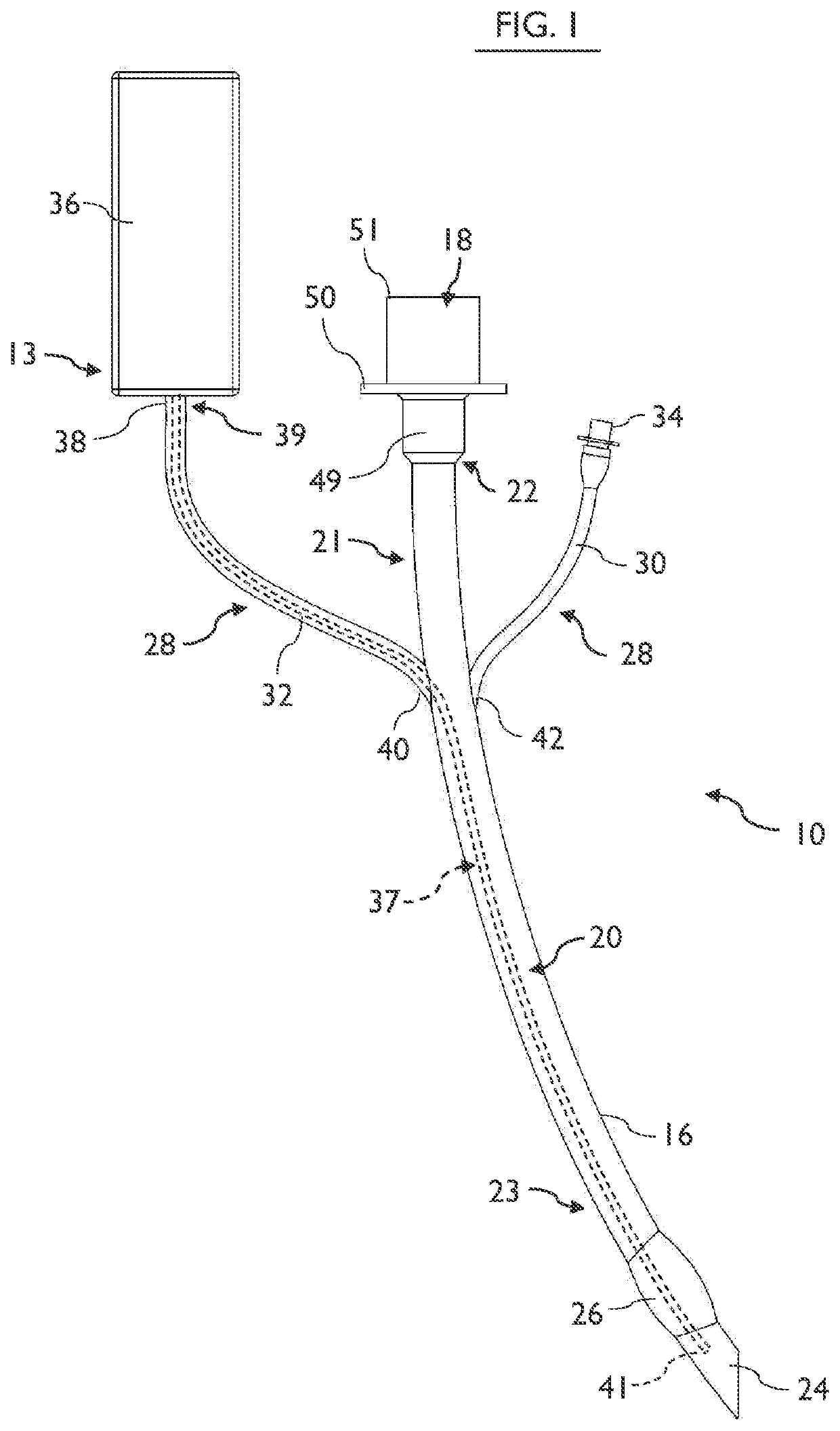
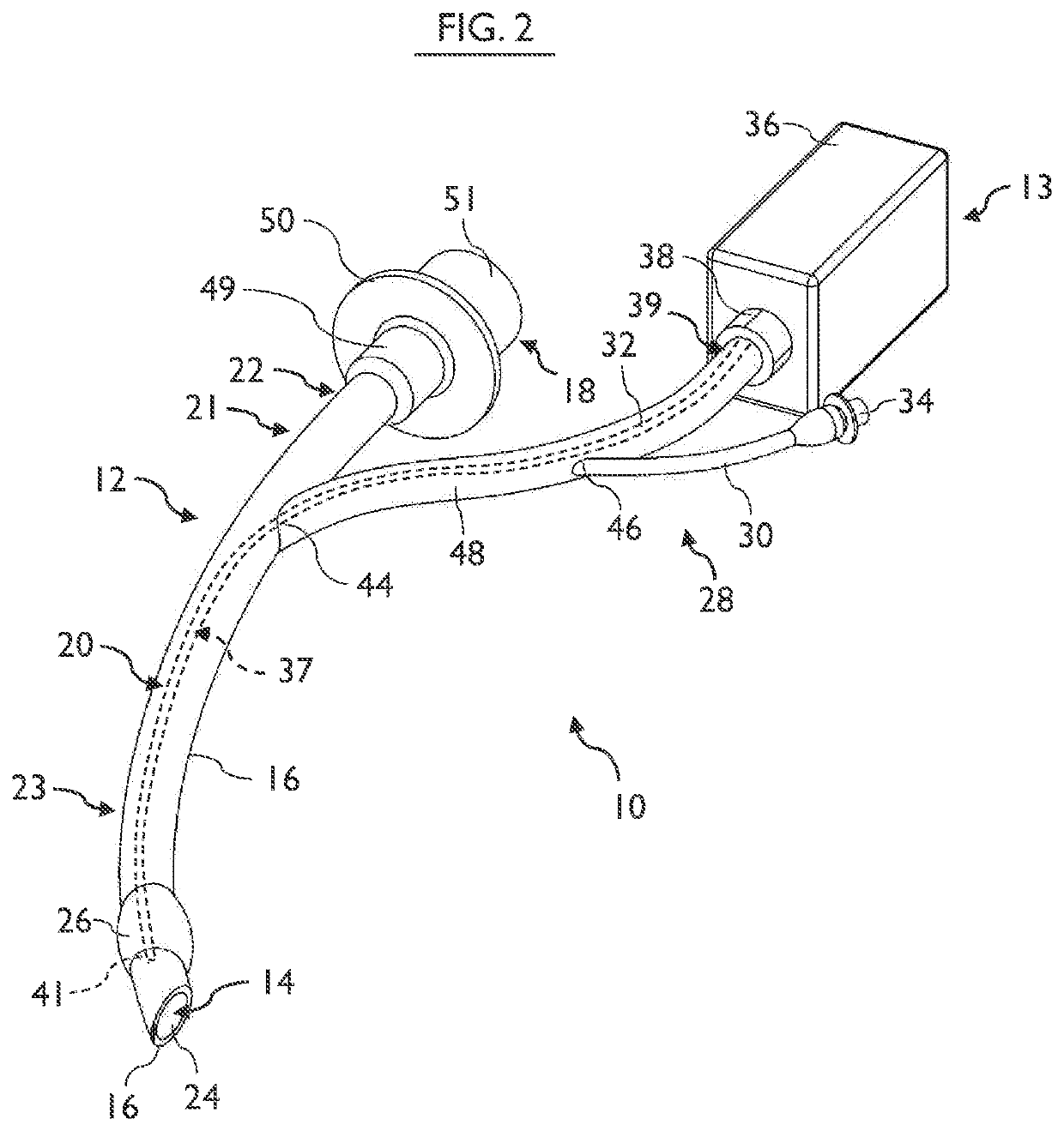
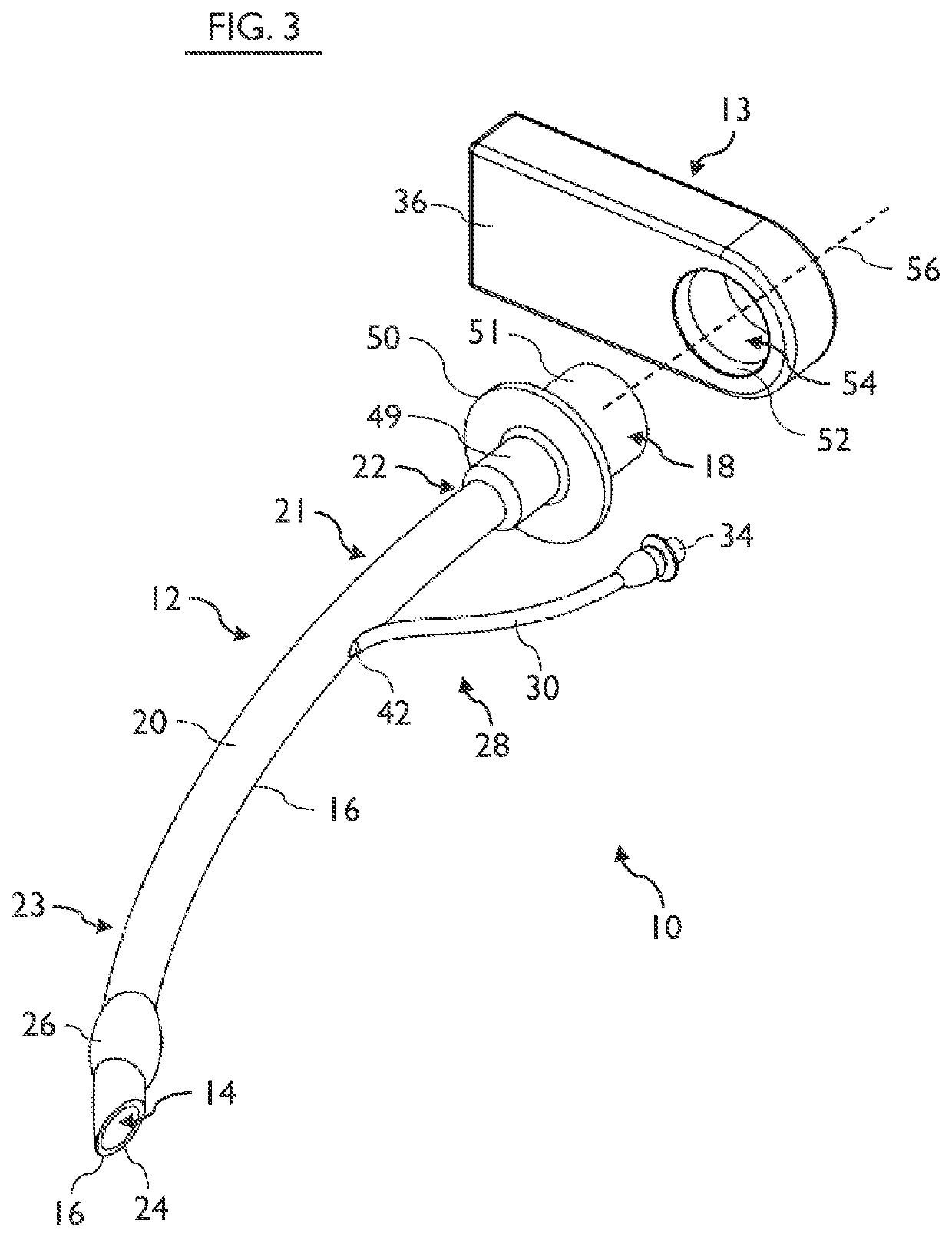
[0012]It should also be understood that the exemplary embodiments of this disclosure include retrofitting a EMR delivery system to an existing standard endotracheal tube where the EMR delivery system is permanently attached to the endotracheal tube or where the EMR delivery system is removably insertable into the endotracheal tube, as well as including the use of a custom-made endotracheal tube that incorporates the EMR delivery system into the structure of the endotracheal tube. For a rapid, retrofit connection of the EMR delivery system onto the endotracheal tube a quick-connect coupling may be used and / or the insertable portion of the EMR delivery system may move freely and axially relative to the endotracheal tube.

Problems solved by technology

Unfortunately, they commonly cause hospital-acquired pneumonia (HAP).
However, establishing the diagnosis of ventilator-associated pneumonia (VAP) can be difficult due to concomitant etiologies and symptoms, leading to increased mortality.
Additionally, the prevalence of drug-resistant bacteria often necessitates a more toxic second-line antibiotic treatment and, due to a greater side-effect profile, further increases the risk to patient safety.
However, these proposals are only marginally effective in-vivo and are unable to prevent numerous infections and deaths.
While possibly helpful for the device insertion, this has not become a standard of care due to simpler methods being developed.
This also does nothing to reduce the infection rates.
Unfortunately, UV light is well known to cause damage to living cells (Riffle.
Unfortunately, these methods and solutions fail to offer a reduction in hospital-acquired infections.
This is due not only to the difficulty in diagnosing HAP in the midst of concomitant etiologies, but also to the high clinical prevalence of drug resistant microorganisms.

Method used


Image

  • Methods and apparatus to deliver therapeutic non-ultraviolet electromagnetic radiation for an endotracheal tube
  • Methods and apparatus to deliver therapeutic non-ultraviolet electromagnetic radiation for an endotracheal tube
  • Methods and apparatus to deliver therapeutic non-ultraviolet electromagnetic radiation for an endotracheal tube

Examples


Embodiment Construction

[0034]Various exemplary embodiments of the present disclosure are described more fully hereafter with reference to the accompanying drawings. These drawings illustrate some, but not all of the embodiments of the present disclosure. It will be readily understood that the components of the exemplary embodiments, as generally described and illustrated in the Figures herein, could be arranged and designed in a wide variety of different configurations. Thus, the following more detailed description of the exemplary embodiments of the apparatus, system, and method of the present disclosure, as represented in FIGS. 1 through 15, is not intended to limit the scope of the invention, as claimed, but is merely representative of exemplary embodiments.

[0035]The phrases “connected to,”“coupled to” and “in communication with” refer to any form of interaction between two or more entities, including mechanical, electrical, magnetic, electromagnetic, fluid, and thermal interaction. Two components may ...


PUM

PropertyMeasurementUnit
wavelengthaaaaaaaaaa
wavelengthsaaaaaaaaaa

Abstract

A therapeutic endotracheal tube assembly is provided for insertion into a patient's trachea to ventilate, to maintain patency of the patient's airway, and to deliver therapeutic electromagnetic radiation (EMR) to the patient. The therapeutic endotracheal tube assembly has an endotracheal tube and an EMR delivery system. The EMR delivery system has an EMR source for emitting non-ultraviolet, therapeutic EMR having intensity sufficient to activate desired therapeutic properties within the patient and an EMR conduction line conducive to the propagation of EMR from the EMR source along the endotracheal tube. The EMR conduction line is removably insertable into the endotracheal tube. The therapeutic endotracheal tube assembly may be custom made or may be constructed by retrofitting a removably insertable EMR delivery system to an existing endotracheal tube.

Description

RELATED APPLICATIONS[0001]This application claims the benefit of U.S. Provisional Application No. 62 / 154,789 that was filed Apr. 30, 2015, for an invention titled METHODS AND APPARATUS TO INACTIVATE INFECTIOUS AGENTS ON AN ENDOTRACHEAL TUBE, which is hereby incorporated in its entirety by this reference. This application also claims the benefit of U.S. Provisional Application No. 62 / 292,028 that was filed Feb. 5, 2016, for an invention titled METHOD AND APPARATUS FOR REMOVABLE CATHETER VISUAL LIGHT STERILIZATION SYSTEM, which is hereby incorporated in its entirety by this reference.[0002]This application is related to a co-pending application entitled METHODS AND APPARATUS TO INACTIVATE INFECTIOUS AGENTS ON A CATHETER RESIDING IN A BODY CAVITY, U.S. application Ser. No. 13 / 801,750, filed Mar. 13, 2013.TECHNICAL FIELD[0003]The present invention is a method and apparatus to provide therapeutic doses of non-ultraviolet light and / or sterilizing doses of non-ultraviolet light to stimulat...

Claims


Application Information

Patent Timeline
Patent Type & Authority Patents(United States)
IPC IPC(8): A61N5/06A61L2/08A61M16/08A61M16/04A61N5/067
CPCA61M16/0816A61N5/0603A61L2/08A61M16/0434A61M16/04A61M16/0486A61M2205/051A61N2005/063A61N2005/0659A61N2005/0663A61N2005/0604A61M16/0833A61N2005/067A61N2005/0651A61M2209/10A61L2/0029A61L2/0058A61L2/0052A61N5/067
Inventor BARNECK, MITCHELL D.RHODES, NATHANIEL L.DE LA PRESA, MARTINPOURSAID, ARASH E.
Owner LIGHT LINE MEDICAL INC
Learn More