Cerebral ischemia diagnosis assisting apparatus, X-ray computer tomography apparatus, and apparatus for aiding diagnosis and treatment of acute cerebral infarct

a technology of cerebral ischemia and assisting equipment, which is applied in the field of cerebral ischemia diagnosis assisting equipment, x-ray computer tomography equipment, and the apparatus for aiding diagnosis and treatment of acute cerebral infarct, can solve the problems of reducing the ct value or the disappearance of the cerebral sulci caused by the ischemia, and the risk of bleeding

Inactive Publication Date: 2005-05-26
KK TOSHIBA +2
View PDF11 Cites 75 Cited by
  • Summary
  • Abstract
  • Description
  • Claims
  • Application Information

AI Technical Summary

Benefits of technology

[0008] It is an object of the invention to assist to read a cere...

Problems solved by technology

However, actually, it is a current state that the reduction in the CT value or the disappearance of the cerebral sulci caused by the ischemia is very delicate and cannot be read without a me...

Method used


Image

  • Cerebral ischemia diagnosis assisting apparatus, X-ray computer tomography apparatus, and apparatus for aiding diagnosis and treatment of acute cerebral infarct
  • Cerebral ischemia diagnosis assisting apparatus, X-ray computer tomography apparatus, and apparatus for aiding diagnosis and treatment of acute cerebral infarct
  • Cerebral ischemia diagnosis assisting apparatus, X-ray computer tomography apparatus, and apparatus for aiding diagnosis and treatment of acute cerebral infarct

Examples


first embodiment

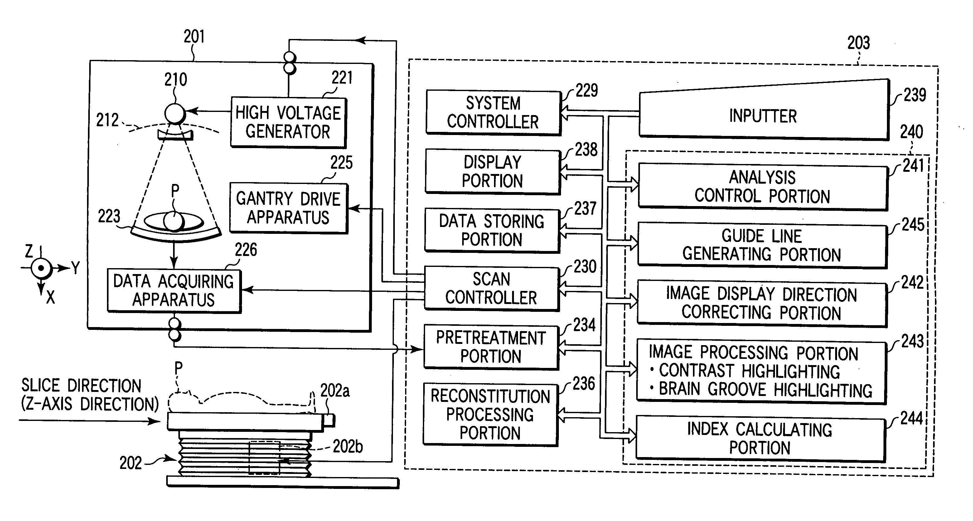
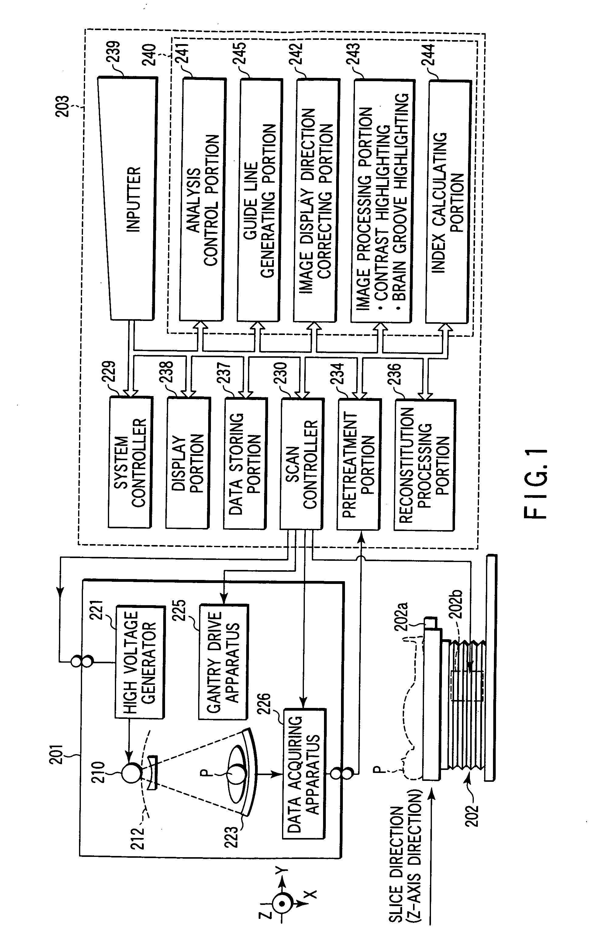
[0052] As shown by FIG. 1, an X-ray computer tomography apparatus includes a gantry 201 constituted for acquiring projected data with regard to a subject. The gantry 201 includes an X-ray tube 210 and an X-ray detector 223. The X-ray tube 210 and the X-ray detector 223 are mounted to a rotating frame 212 in a ring-like shape driven to rotate by a gantry drive apparatus 225. A central portion of the rotating frame 212 is opened and a subject P mounted on a patient couch top 202a of a patient couch 202 is inserted into the opening portion. Further, a rotational center axis of the rotating frame 212 is defined by Z-axis (slice direction axis) and a plane orthogonal to Z-axis is defined by orthogonal two axes of X and Y.

[0053] A tube voltage is applied from a high voltage generator 221 between a cathode and an anode of the X-ray tube 210 and a filament current is supplied to a filament of the X-ray tube 210 from the high voltage generator 221. X-ray is generated by applying the tube vo...

second embodiment

[0106] An apparatus for aiding the diagnosis and treatment of acute cerebral infarction is described below by referring to the drawings, the apparatus being built according to the present invention. This aiding apparatus for the diagnosis and treatment of acute cerebral infarction treats CT images (spatial distribution of CT values) obtained by an X-ray computed tomography system (CT scanner). In the description given below, it is assumed that the aiding apparatus for the diagnosis and treatment of acute cerebral infarction is incorporated within the X-ray CT scanner. The aiding apparatus for the diagnosis and treatment of acute cerebral infarction can also be constructed separately from the X-ray CT scanner as a standalone apparatus.

[0107] As shown in FIG. 13, the X-ray CT scanner according to the present aspect has a gantry 1 designed to collect projection data about a patent to be examined. The gantry 1 has an X-ray tube 10 and an X-ray detector 23, which are mounted on an annul...

third embodiment

[0182] An apparatus for aiding the diagnosis and treatment of acute cerebral infarction, according to the third embodiment of the invention, will be described with reference to the drawings. The apparatus according to this embodiment uses a three-dimensional CT image that an X-ray CT scanner has acquired. The three-dimensional CT image is of either multi-slice type or volume type that is a set of voxels. Here, the CT image will be described as a multi-slice type one.

[0183] As seen from FIG. 31, the X-ray CT scanner used in the present embodiment is similar to its counterpart of the second-embodiment. It differs in two respects. First, the image processing portion 43 can extract images of the blood vessels. Second, the apparatus further comprises a 3D-image processing portion 51.

[0184] As FIGS. 32 and 33 show, simple CT scanning is performed, acquiring projection data representing a 3-dimensional region of the patient's head (S1). Contrast dynamic CT scanning is also performed in s...


PUM


Abstract

A cerebral ischemia diagnosis assisting apparatus comprises a storing portion for storing multi-slices or volume data with regard to the head portion of a subject, an image generating portion for generating a tomography image of a brain from the multi-slices or the volume data, an image processing portion for processing the tomography image for generating a contrast highlighting image and a brain sulci highlighting image or either thereof, and a display portion for displaying the tomography image along with the contrast highlighting image and the brain sulci highlighting image.

Description

CROSS-REFERENCE TO RELATED APPLICATIONS [0001] This application is based upon and claims the benefit of priority from prior Japanese Patent Applications No. 2003-368893, filed Oct. 29, 2003; and No. 2003-368894, filed Oct. 29, 2003, the entire contents of both of which are incorporated herein by reference. BACKGROUND OF THE INVENTION [0002] 1. Field of the Invention [0003] The present invention relates to a cerebral ischemia diagnosis assisting apparatus, an X-ray computer tomography apparatus equipped with a function thereof, and an apparatus for aiding the diagnosis and treatment of acute cerebral infarct by offering information useful for diagnosis and determination of treatment policy of acute cerebral infarction. [0004] 2. Description of the Related Art [0005] It is very important in the medical field to qualitatively determine a position and a size of an ischemia region and a degree of seriousness thereof by reading an X-ray CT image of the brain in acute cerebral infarction a...

Claims


Application Information

Patent Timeline
IPC IPC(8): A61B5/05A61B6/03G06T5/00G06T7/00
CPCA61B6/481A61B6/487A61B6/501A61B6/504G06T7/0012G06T11/001G06T2207/30016A61B6/507G06T7/0081G06T7/608G06T2207/10081G06T7/11G06T7/68
Inventor IKEDA, YOSHIHIROMURPHY, KIERANOKUMURA, MIWAFUJISAWA, YASUKOTOKI, YUUSUKENOSHI, YASUHIRO
Owner KK TOSHIBA
Why Eureka
Try Eureka