Guidewire for crossing occlusions or stenoses

a technology of stenoses and guidewires, applied in the field of medical devices, kits, methods, can solve the problems of myocardial infarction or heart attack, initial guidewire placement is difficult or impossible in tortuous regions of the vasculature, and is equally difficult. to achieve the effect of facilitating passing and better control

Inactive Publication Date: 2005-05-26
REVASCULAR THERAPEUTICS
View PDF91 Cites 71 Cited by
  • Summary
  • Abstract
  • Description
  • Claims
  • Application Information

AI Technical Summary

Benefits of technology

[0007] The present invention provides systems and methods for removing occlusive material and passing through occlusions, stenosis, thrombus, and other material in a body lumen. More particularly, the present invention can be used passing through stenosis or occlusions in a neuro, cardio, and peripheral body lumens. Generally, the present invention includes an elongate member that is positioned adjacent the occlusion or stenosis. A drive shaft having a distal tip is rotated and advanced from within the elongate member to create a path forward of the elongate member to form a path in the occlusion or stenosis. To facilitate passing through the occlusion or stenosis, the distal end of the elongate member can be steerable to provide better control the creation of the path through the occlusion or stenosis. Optionally, the target site can be infused and / or aspirated before, during, and after creation of the path through the occlusion.
[0008] In an exemplary embodiment, the elongate member is a hollow guidewire that has a flexibility, pushability and torqueability to be advanced through the tortuous blood vessel without the use of a separate guidewire. Additionally, the hollow guidewire may be sized to fit within a conventional support or access catheter system and inserted into the blood vessel and delivered to the target site. The catheter system can be delivered either concurrently with the advancement of the hollow guidewire or after the guidewire has reached the target site. The position of the hollow guidewire and catheter system can be maintained and stabilized while the drive shaft is rotated and translated out of the axial lumen of the hollow guidewire. The distal tip of the drive shaft can be deflected, coiled, blunted, flattened, enlarged, twisted, basket shaped, or the like. In some embodiments, to increase the rate of removal of the occlusive material, the distal tip is sharpened or impregnated with an abrasive material such as diamond chips, diamond powder, glass, or the like.
[0010] In a specific configuration, the drive shaft has spiral threads or external riflings extending along the shaft. The spirals typically extend from the proximal end of the shaft to a point proximal of the distal tip. As the drive shaft is rotated and axially advanced into the occlusive material, the distal tip creates a path and removes the material from the body. The rotating spirals act similar to an “Archimedes Screw” and transport the removed material proximally up the lumen of the elongate member and prevent the loose atheromatous material from escaping into the blood stream.
[0012] In other embodiments, a hollow guidewire can be used as the support or access system. The hollow guidewire can be navigated to and positioned at the target site, with or without the use of a separate guidewire. The hollow guidewire support system provides the flexibility, maneuverability, torqueability (usually 1:1), and columnar strength necessary for accurately advancing through the tortuous vasculature. The hollow guidewire support system can act as a working channel inside of which other interventional devices can be delivered to the target site. Such devices include, but are not limited to a rotating guidewire, infusion guidewire, clot maceration guidewire, normal guidewire, and the like. Because the hollow guidewire is not composed of polymer, the hollow guidewire working channel does not soften at body temperatures.
[0013] The hollow guidewire working channel typically has a thin wall construction which allows the lumen of the working channel to be maximized when compared with polymeric based catheter designs. This allows larger diameter devices to be inserted into it than can be inserted through similar sized catheter-based devices. The larger lumen of the hollow guidewire working channel allows devices such as clot macerators and other larger devices to be delivered to the target lesion. Additionally the larger diameter lumen allows infusion or clot dissolving fluid and / or aspiration of the debris created in the clot maceration process.

Problems solved by technology

Atheromatous and other vascular deposits restrict blood flow and can cause ischemia which, in acute cases, can result in myocardial infarction or a heart attack.
Initial guidewire placement, however, can be difficult or impossible in tortuous regions of the vasculature.
Moreover, it can be equally difficult if the lesion is total or near total, i.e. the lesion occludes the blood vessel lumen to such an extent that the guidewire cannot be advanced across.
While effective in some cases, these catheter systems, even with a separate guidewire, have great difficulty in traversing through the small and tortuous body lumens of the patients and reaching the target site.

Method used


Image

  • Guidewire for crossing occlusions or stenoses
  • Guidewire for crossing occlusions or stenoses
  • Guidewire for crossing occlusions or stenoses

Examples


Embodiment Construction

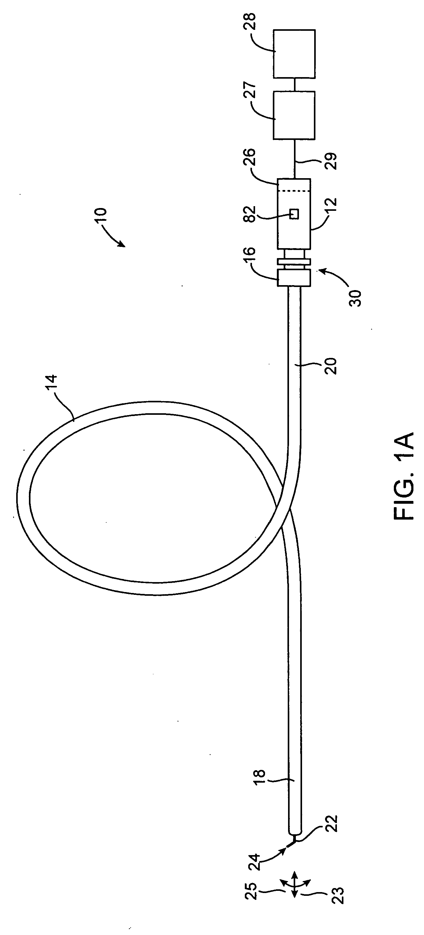
[0050] The systems, devices and methods according to the present invention will generally be adapted for the intraluminal treatment of a target site within a body lumen of a patient, usually in a coronary artery or peripheral blood vessel which is occluded or stenosed with atherosclerotic, stenotic, thrombotic, or other occlusive material. The systems, devices and methods, however, are also suitable for treating stenoses of the body lumens and other hyperplastic and neoplastic conditions in other body lumens, such as the ureter, the biliary duct, respiratory passages, the pancreatic duct, the lymphatic duct, and the like. Neoplastic cell growth will often occur as a result of a tumor surrounding and intruding into a body lumen. Removal of such material can thus be beneficial to maintain patency of the body lumen. While the remaining discussion is directed at passing through atheromatous or thrombotic occlusive material in a coronary artery, it will be appreciated that the systems an...


PUM


Abstract

Systems and methods for crossing stenosis, partial occlusions, or complete occlusions within a body lumen. The systems generally include an elongate member such as a hollow guidewire that houses a rotatable and translatable drive shaft. The drive shaft typically has a distal portion that is advanced to create a path in the occlusive material that is large enough to allow the hollow guidewire to cross the occlusive material.

Description

CROSS-REFERENCES TO RELATED APPLICATIONS [0001] The present application claims benefit of U.S. Patent Application No. 60 / 195,154, filed Apr. 6, 2000, under 37 C.F.R. §1.78, the complete disclosure of which is incorporated herein by reference. The present application is also related to U.S. patent application Ser. No. 09 / 030,657, filed Feb. 25, 1998, the complete disclosure of which is herein incorporated by reference.BACKGROUND OF THE INVENTION [0002] The present invention is generally related to medical devices, kits, and methods. More specifically, the present invention provides a system for crossing stenosis, partial occlusions, or total occlusions in a patient's body. [0003] Cardiovascular disease frequently arises from the accumulation of atheromatous material on the inner walls of vascular lumens, particularly arterial lumens of the coronary and other vasculature, resulting in a condition known as atherosclerosis. Atheromatous and other vascular deposits restrict blood flow an...

Claims


Application Information

Patent Timeline
Patent Type & Authority Applications(United States)
IPC IPC(8): A61B17/22A61B17/32
CPCA61B17/320758A61B2017/22042A61B2017/22044A61B2017/22069A61B17/50A61B2017/320004A61B17/3207A61B17/3421A61B2017/22077
Inventor NORIEGA, GERARDO V.CHECHELSKI, VICTORSUDARIA, RUDOLFO
Owner REVASCULAR THERAPEUTICS
Why Eureka
Try Eureka