
Hierarchical medical image view determination

Inactive Publication Date: 2006-03-23
SIEMENS MEDICAL SOLUTIONS USA INC
View PDF10 Cites 59 Cited by
  • Summary
  • Abstract
  • Description
  • Claims
  • Application Information

AI Technical Summary

Benefits of technology

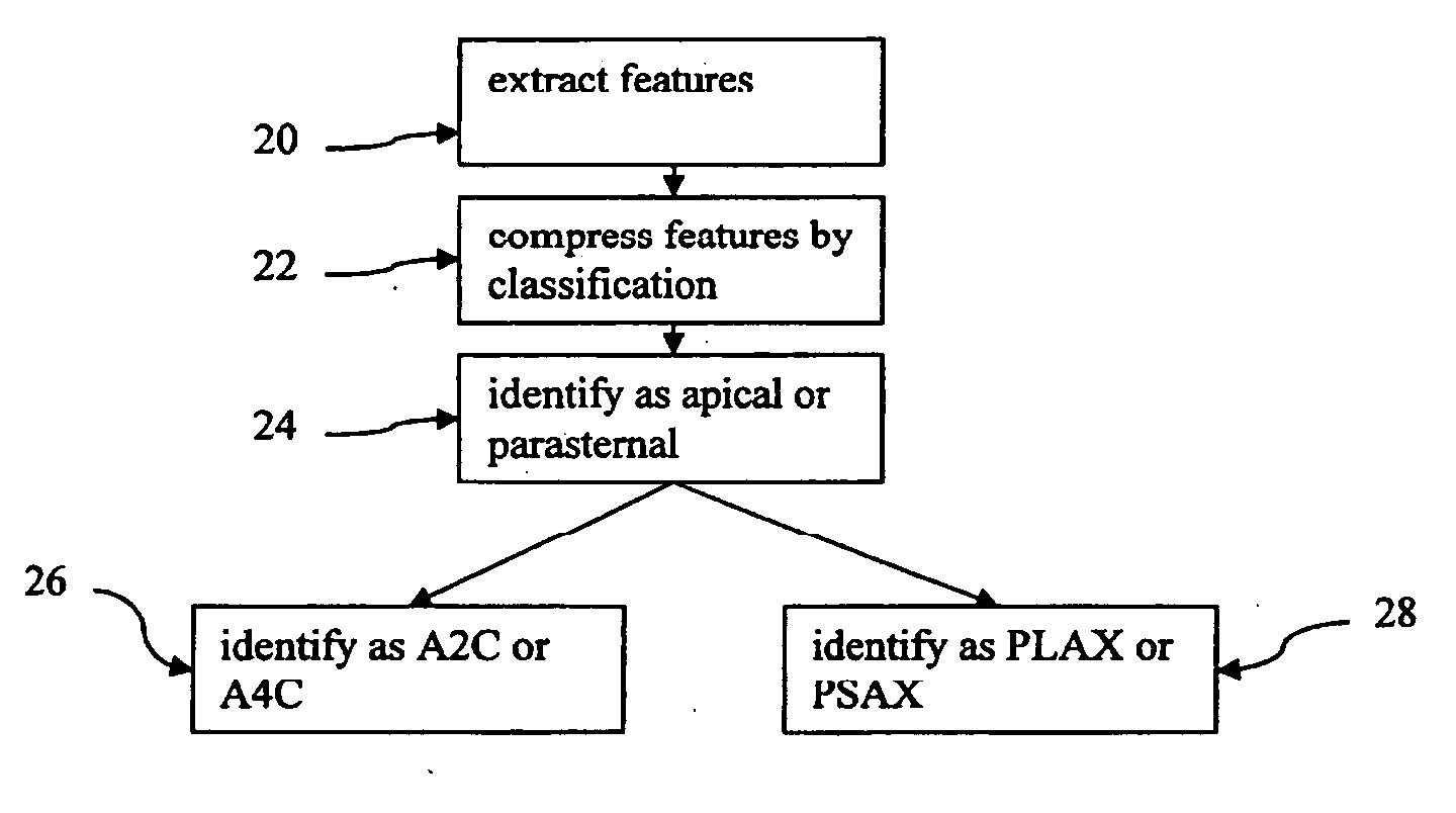
[0007] In a first aspect, a method is provided for identifying a cardiac view of a medical ultrasound image. With a processor, the medical ultrasound image is classified between any two or more of parasternal, apical, subcostal, suprasternal or unknown. With the processor, the cardiac view of the medical image is classified as a particular parasternal or apical view based on the classification as parasternal or apical, respectively.
[0008] In a second aspect, a system is provided for identifying a cardiac view of a medical ultrasound image. A memory is operable to store medical ultrasound data associated with the medical ultrasound image. A processor is operable to classify the medical ultrasound image between any two or more of subcostal, suprasternal, unknown, parasternal or apical from the medical ultrasound data, and is operable to classify the cardiac view of the medical image as a particular parasternal or apical view based on the classification as parasternal or apical, respectively.
[0009] In a third aspect, a computer readable storage media has stored therein data representing instructions executable by a programm

Problems solved by technology

Depending on the skill and knowledge of the reviewing physician, clinician, or radiologist, manual evaluation of medical images can result in misdiagnosed medical conditions due to simple human error.
Furthermore, when the acquired medical im

Method used


Image

  • Hierarchical medical image view determination
  • Hierarchical medical image view determination
  • Hierarchical medical image view determination

Examples


Example

DETAILED DESCRIPTION OF THE DRAWINGS AND PRESENTLY PREFERRED EMBODIMENTS

[0020] Ultrasound images of the heart can be taken from many different angles. Efficient analysis of these images requires recognizing which position the heart is in so that cardiac structures can be identified. Four standard views include the apical two-chamber view, the apical four-chamber view, the parasternal long axis view, and the parasternal short axis view. Other views or windows include: apical five-chamber, parasternal long axis of the left ventricle, parasternal long axis of the right ventricle, parasternal long axis of the right ventricular outflow tract, parasternal short axis of the aortic valve, parasternal short axis of the mitral valve, parasternal short axis of the left ventricle, parasternal short axis of the cardiac apex, subcostal four chamber, subcostal long axis of inferior vena cava, suprasternal north long axis of the aorta, and suprasternal notch short axis of the aortic arch. To assis...


PUM


Abstract

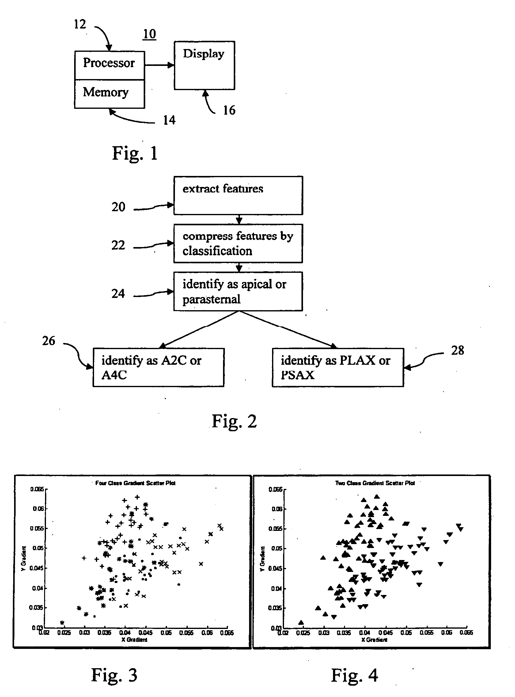
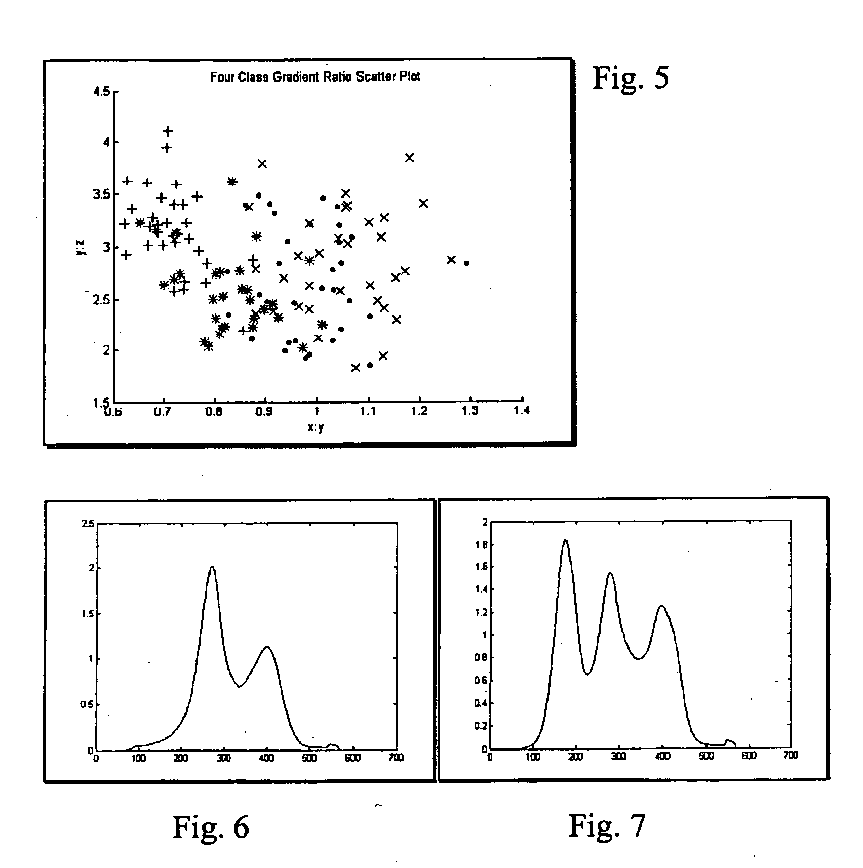
A cardiac view of a medical ultrasound image is automatically identified. By grouping different views into sub-categories, a hierarchal classifier identifies the views. For example, apical views are distinguished from parasternal views. Specific types of apical or parasternal views are identified based on distinguishing between images of the geneses. Different features are used for classifying, such as gradients, functions of the gradients, statistics of an average frame of data from a clip or sequence of frames, or a number of edges along a given direction. The number of features used may be compressed, such as by classifying a plurality of features into a new feature. For example, alpha weights in a model of features and classes are determined and used as features for classification.

Description

RELATED APPLICATIONS [0001] The present patent document claims the benefit of the filing date under 35 U.S.C. §119(e) of Provisional U.S. Patent Application Ser. No. 60 / 611,865, filed Sep. 21, 2004, the disclosure of which is hereby incorporated by reference.BACKGROUND [0002] The present invention relates to classifying medical images. For example, a processor identifies cardiac views associated with medical ultrasound images. [0003] In the field of medical imaging, various imaging modalities and systems generate medical images of anatomical structures of individuals for screening and evaluating medical conditions. These imaging systems include, for example, CT (computed tomography) imaging, MRI (magnetic resonance imaging), NM (nuclear magnetic) resonance imaging, X-ray systems, US (ultrasound) systems, PET (positron emission tomography) systems, or other systems. With ultrasound, sound waves propagate from a transducer towards a specific part of the body (the heart, for example). ...

Claims


Application Information

Patent Timeline
IPC IPC(8): A61B8/02
CPCG06K9/6217G06T2207/30048G06T7/0012G06K9/6282G06F18/21G06F18/24323
Inventor KRISHNAN, SRIRAMBI, JINBORAO, R. BHARATSTOECKEL, JONATHANOTEY, MATTHEW ERIC
Owner SIEMENS MEDICAL SOLUTIONS USA INC
Learn More