Methods and sensors for monitoring internal tissue conditions

a technology of internal tissue and sensors, applied in the field of methods and sensors for monitoring internal tissue conditions, can solve the problems of affecting the continuous monitoring of organ condition, affecting the effect of oxygen circulation,

Inactive Publication Date: 2006-09-07
MANSOUR HEBAH NOSHY +1
View PDF13 Cites 50 Cited by
  • Summary
  • Abstract
  • Description
  • Claims
  • Application Information

AI Technical Summary

Problems solved by technology

Surgical complications, such as vascular complications, may disrupt adequate oxygen circulation to the tissue, which is critical to organ function and survival.
Unfortunately, these blood tests reflect liver condition only at the time the blood sample is drawn, and changes in these laboratory values can often be detected only after significant organ damage has already occurred, permitting a limited opportu

Method used


Image

  • Methods and sensors for monitoring internal tissue conditions
  • Methods and sensors for monitoring internal tissue conditions
  • Methods and sensors for monitoring internal tissue conditions

Examples


Embodiment Construction

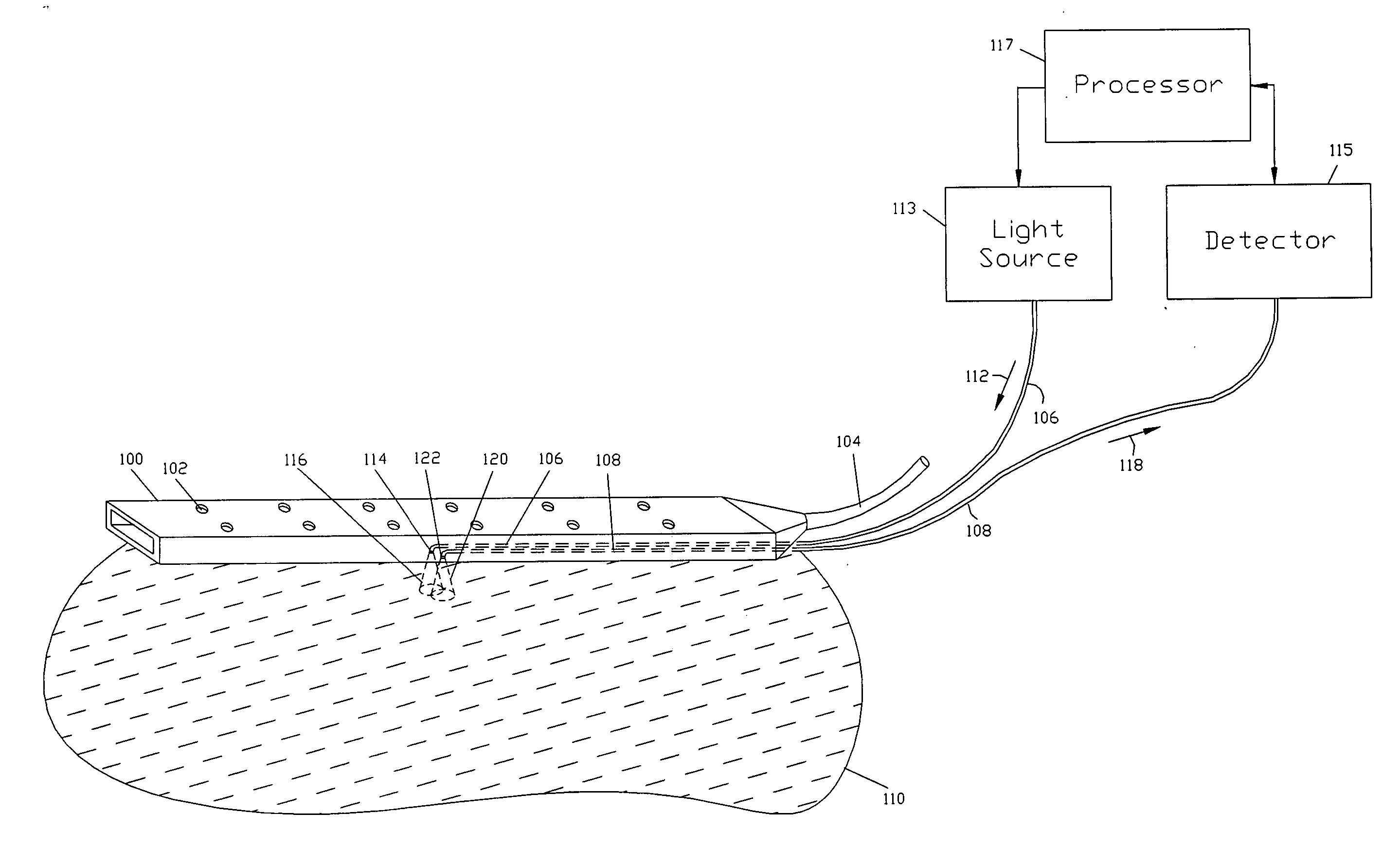
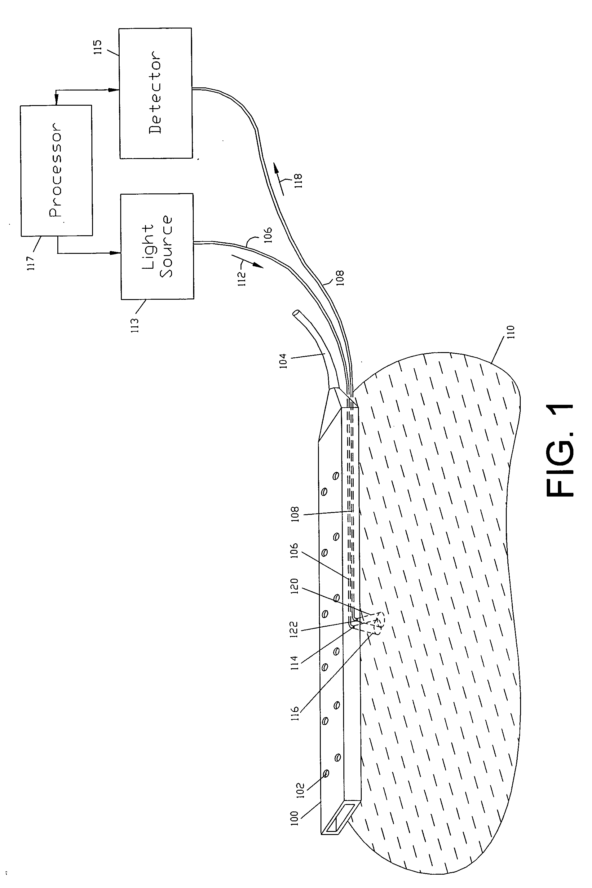
[0034] Referring to FIG. 1, the present invention includes a surgical drain 100 with draining holes 102, a drainage collection tube 104, a first optical fiber 106, and a second optical fiber 108. The drain 100 is placed on the tissue 110 during surgery and the optical fibers 106 and 108 may be used to optically interrogate the tissue 110 to determine its condition.

[0035] The first optical fiber 106 may transmit the excitation radiation 112 that emerges from the distal aperture 114 of the optical fiber 106 to irradiate the cone of acceptance 116 within the tissue 110 and generate a returned radiation. The excitation radiation 112 may be generated by the light source 113. The light source 113 may be a lamp, a light emitting diode (LED), a combination of light emitting diodes of different wavelengths, or a laser. The excitation radiation 112 may be preferably in the visible wavelength band between 450 and 600-nm, however, it may be also in the ultraviolet, and / or infrared wavelength b...


PUM


Abstract

The present invention relates to fiberoptic sensors embedded within a surgical drain to monitor the condition of tissue proximate to the surgical drain and methods of processing data from the sensors. The light transmitting and collecting tips of the optical fibers are adapted to enable sideways interrogation of the adjacent tissues using optical techniques to measure the tissue condition. The light transmitting and collecting tips of the optical fibers may be bent at angle, disposed between a light absorbing and transmitting sheets, or uncladded to enable sideways emission and collection of light. Processing methods for reducing the site dependency of the measured data, maximizing the signal to noise ratio, and increasing the measurement sensitivity to changes in the tissue condition are described.

Description

CROSS REFERENCE TO RELATED APPLICATIONS [0001] This application claims the benefit of U.S. Provisional Application No. 60 / 652,939, filed Feb. 14, 2005, entitled Methods and Sensors for Monitoring Internal Tissue Conditions, the entire contents of which application are incorporated herein by reference.FIELD OF THE INVENTION [0002] The present invention relates to optical sensors embedded within a surgical drain to monitor the condition of organs and tissues proximate to the surgical drain and methods of processing data from the sensors. BACKGROUND OF THE INVENTION [0003] Monitoring of organ condition may be important after surgery such as organ transplantation and resection. Surgical complications, such as vascular complications, may disrupt adequate oxygen circulation to the tissue, which is critical to organ function and survival. Following liver surgery, for example, a physician may draw patient blood to determine the condition of the organ by measuring liver enzymes (such as tran...

Claims


Application Information

Patent Timeline
IPC IPC(8): A61B5/00
CPCA61B5/0059A61B5/14539A61B5/413
Inventor MANSOUR, HEBAH NOSHYSHEHADA, RAMEZ EMILE NECOLA
Owner MANSOUR HEBAH NOSHY
Why Eureka
Try Eureka