Device and method for recording and representing images of a test object

a technology for test objects and devices, applied in medical science, othalmoscopes, diagnostics, etc., can solve problems such as rendering measurements useless, and achieve the effects of reducing light stress, simple, practicable and extremely economical construction variants

Active Publication Date: 2007-08-02
IMEDOS INTELLIGENTE OPTISCHE SYST DER MEDIZIN & MESSTECHNIK GMBH
View PDF10 Cites 6 Cited by
  • Summary
  • Abstract
  • Description
  • Claims
  • Application Information

AI Technical Summary

Benefits of technology

[0010] It is the primary object of the invention to record images of the object to be examined with reduced light stress and lower expenditure on adjustments and to generate secondary images which are substantially independent from brightness, are highly suitable for spectrometric studies of the metabolism and microcirculation in the eye as well as for functional imaging, and can be adapted to the medical inquiry and make it possible to provide complex secondary image information while also enabling simple, practicable and extremely economical constructional variants.

Problems solved by technology

Due to different, uncomparable illumination situations within an image (varying spatial lighting) and between different images, e.g., a change in the main focus of illumination, occurrence of shadows or reflections during spontaneous eye movement, the determination of very slight changes in brightness, position and shape that is required for purposes of detection is subject to a high error rate which sometimes renders measurements useless.
This optical concept is disadvantageous in particular because of the adjustments required for implementing the partial beam paths, the extremely high light stress to which the patient is subjected, and the fact that no simple constructional variants which would simplify the apparatus and reduce costs can be derived from the concept.

Method used


Image

  • Device and method for recording and representing images of a test object
  • Device and method for recording and representing images of a test object
  • Device and method for recording and representing images of a test object

Examples


Embodiment Construction

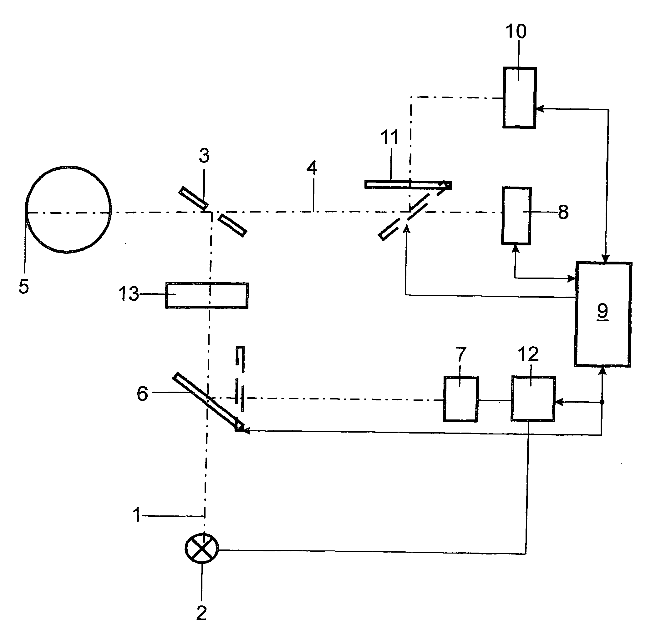
[0057] The first embodiment example shows a simple and distinctly economical construction according to the invention for displaying blood volume independent from brightness. The arrangement comprises the elements of any retina camera, wherein a filter 13 according to the invention which is spectrally adapted according to the invention to an electronic color camera 8 as will be described in the following is arranged in a common illumination beam path 1 of an illumination system containing at least one illumination source 2. The electronic images are supplied to a controlling and evaluating unit, e.g., a controlling and evaluating computer 9, which serves to generate and display secondary images and functional images and to store them advantageously in a patient-specific manner. The other elements in FIG. 1 which form the illumination beam path 1 and the recording beam path 4 are known from retina camera technology. The elements include a perforated mirror 3, a recording beam path 4 p...


PUM


Abstract

It is the object of an arrangement and a method for the recording and reproduction of images of an object to be examined to record the images of the object to be examined with reduced light stress and lower expenditure on adjustments and to generate secondary images which are substantially independent from brightness, are highly suitable for spectrometric studies of metabolism and microcirculation at the eye as well as for functional imaging, and can be adapted to the medical inquiry and make it possible to provide complex secondary image information while also enabling simple, practicable and extremely economical constructional variants. An illumination system contains at least one beam path with a device for simultaneous illumination of the object to be examined by at least one reference wavelength region and at least one information wavelength region, each of which is adapted, respectively, to a color channel of an image-generating recording system. While the at least one reference wavelength region is at least approximately invariant with respect to medically relevant information, the at least one information wavelength region is provided for detecting the medically relevant information. The method according to the invention combines the image values of evaluation windows or individual image points of simultaneously recorded images to form secondary images and image sequences while generating spatially-resolved dynamic characteristic values which are combined to form functional images.

Description

CROSS-REFERENCE TO RELATED APPLICATIONS [0001] This application claims priority of International Application No. PCT / DE2005 / 000287, filed Feb. 16, 2005 and German Application No. 10 2004 008 675.3, filed Feb. 20, 2004, the complete disclosures of which are hereby incorporated by reference.BACKGROUND OF THE INVENTION [0002] a) Field of the Invention [0003] The invention is directed to an arrangement for recording and reproducing images of an object to be examined having an illumination system, an image-generating recording system, and a controlling and evaluating computer. The arrangement is suitable in particular for measurement systems for spectrometric examination of metabolism and of microcirculation which graphically display spatial or temporal differences in order to determine pathological changes or therapeutic changes in the object to be examined. [0004] Further, the invention is directed to an image-generating method for detecting spatial and / or temporal, medically relevant ...

Claims


Application Information

Patent Timeline
Patent Type & Authority Applications(United States)
IPC IPC(8): A61B6/00A61B3/12A61B5/12
CPCA61B5/12
Inventor VILSER, WALTHARDSEIFERT, BERND ULLRICHRIEMER, THOMASFINK, AXEL
Owner IMEDOS INTELLIGENTE OPTISCHE SYST DER MEDIZIN & MESSTECHNIK GMBH
Why Eureka
Try Eureka