Brain Function Scan System

a function scan and brain technology, applied in the field of brain function scan system, can solve the problems of difficult for emergency personnel to determine if a subject is suitable, inability to detect, and inability to provide optimal treatmen

Inactive Publication Date: 2009-09-10
NEW YORK UNIV
View PDF7 Cites 25 Cited by
  • Summary
  • Abstract
  • Description
  • Claims
  • Application Information

AI Technical Summary

Benefits of technology

[0017] In accordance with the present invention, there is provided a portable EEG device which can accurately, reliably, continuously and quickly determine if the patient is in a coma, is suffering from concussion or is brain dead; if he is having an ischemic stroke or an intracerebral hemorrhage; if he has a serious spinal injury; and, if his behavior is of concern, whether he has abnormal brain function.
[0019] Preferably, stimulations are used in two different modes, i.e., auditory clicks and electric pulses to the skin. The stimuli, although concurrent, are at different prime number frequencies to permit separation of different EPs and avoid interference. Such concurrent stimulations for EP permit a more rapid, and less costly, examination and provide the patient's responses more quickly, which is important in emergency situations. Power spectra of spontaneous EEG, waveshapes of Averaged Evoked Potentials, and extracted measures, such as frequency specific power ratios, can be transmitted to a remote receiver. The latencies of successive EP peaks of the patient may be compared to those of a normal group by use of a normative template.
[0027] One advantage of the portable EEG device is that it allows personnel at the scene of an emergency to determine almost instantly whether a person is alive but suffering from concussion; is having an ischemic stroke or is suffering intracerebral bleeding; if the person is dead or in coma; whether the patient has brainstem or spinal injury; or if the patient does not have a heartbeat. The device may be utilized by medical personnel in the field, ambulance medical personnel, firemen and policemen as well as other emergency room personnel, and may be relatively simple to use and low in cost. The instrument, when used in emergency vehicles, such as firetrucks and ambulances, preferably has a built-in cellular telephone which automatically dials-up or otherwise transmits its data to a neurometric computer, for example, at a hospital. Thus, while providing immediate automatic evaluation of patient's brain state at the emergency site, it can transmit a series of brain measurements which are continuously updated to construct a “state trajectory” for remote evaluation by qualified specialists.

Problems solved by technology

At the present time it is difficult for emergency personnel to determine if a subject has suffered injury to the brain or the spinal cord, cerebrovascular obstruction (stroke) or hemorrhage (bleeding).
Consequently, although the patient may be suffering from brain damage or dysfunction, such as a tumor, it is often not detected in the course of the medical examination.
The absence of information about central nervous system (CNS) dysfunction often results in suboptimal treatment.
Unless the patient is treated promptly, brain cells in the ischemic region would continue to be deprived of oxygen, possibly destroying parts of his cognitive abilities, memory and motor skills and possibly resulting in death.
However, tPA treatment of a possible stroke victim may be hazardous to initiate, as his physical symptoms may be caused by an intracerebral hemorrhage which can be worsened by dissolving clots.
But the ambulance personnel cannot determine if he has suffered spinal injury by simply looking at the comatose patient.
That instrument is large, non-portable and relatively expensive (tens of thousands of dollars).
It is not suitable for use in an ambulance, emergency room or a doctor's office for regular medical examinations.
However, in some emergencies, medical personnel need to quickly determine if a patient has had a stroke or if the patient is alive but in a coma, or dead, or if a person has suffered spinal injury.
A particular difficulty arises when some patients have spinal injury and are unconscious.
In those cases, it would be difficult for medical personnel to ascertain who can safely be moved or should not be moved because of spinal injury.
Persons with such conditions may die due to the lack of medical information, for example, a non-spinal injury patient may be in a coma and is not properly and timely transported to a hospital, or may become paralyzed if moved with unrecognized spinal injury.

Method used


Image

  • Brain Function Scan System
  • Brain Function Scan System
  • Brain Function Scan System

Examples


Embodiment Construction

Version 1

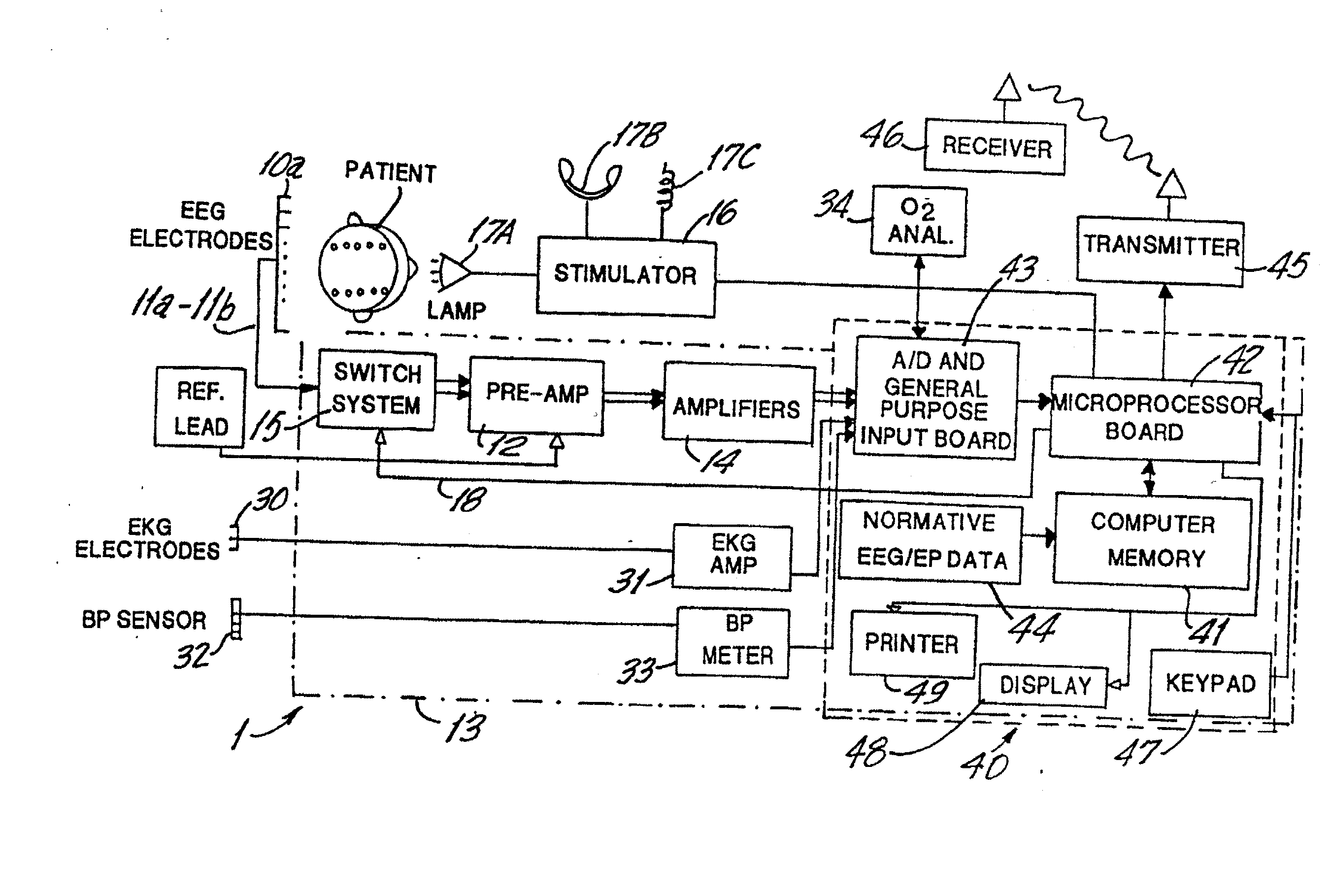
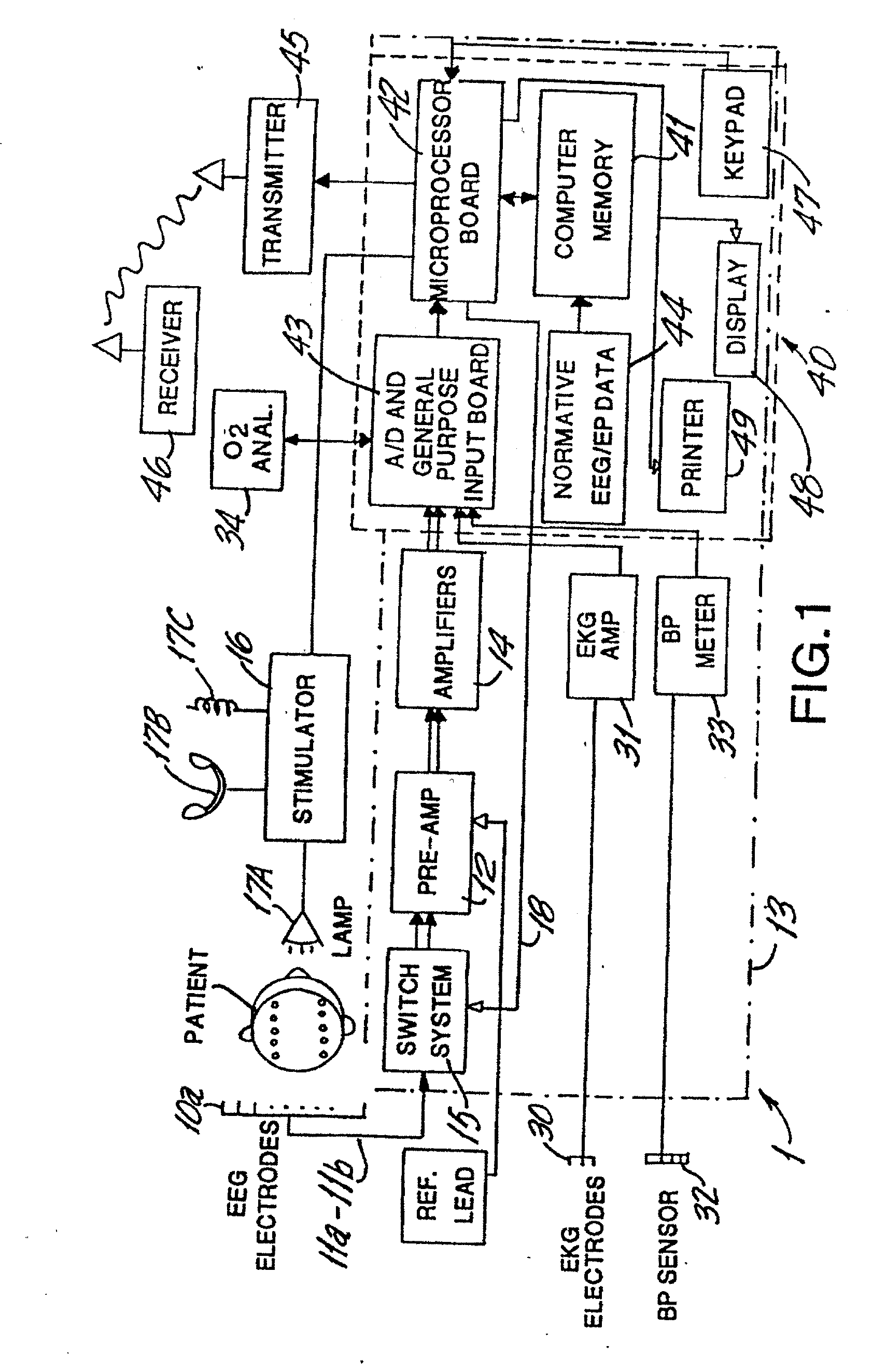
[0035] In the first embodiment, shown in FIG. 1, the instrument 1 is a small and inexpensive device which is portable and may be hand-held. It uses a computer system based on a conventional microprocessor, such as an Intel Pentium I™ and has a limited internal memory, for example, 100 MB.

[0036] The instrument 1 has 1-24 EEG amplifiers, each of which may be connected to a removable EEG electrode shown as electrodes 10a-10p. A suitable electrode uses an adhesive cover, which is removed before applying it to the scalp. The electrode may have multiple small barbs, a needle electrode or a conductive disk, which is removably attached to and may penetrate the patient's skin; the electrode may also use conductive gel, providing rapid attachment and acceptably low impedance, and may be sterile and disposable. In this, and other embodiments, a self-adhering electrode may be used, for example, the “ZIP-PREP”™ electrode having stainless steel micro-barbs in an adhesive gel patch, th...


PUM


Abstract

A portable EEG (electroencephalograph) instrument, especially for use in emergencies and brain assessments in physicians' offices, detects and amplifies brain waves and converts them into digital data for analysis by comparison with data from normal groups. In one embodiment, the EEG electrodes are in a headband which broadcasts the data, by radio or cellular phone, to a local receiver for re-transmission and / or analysis. In another embodiment, the subject is stimulated in two modes, i.e., aural and sensory, at two different frequencies to provide the subject's EPs (Evoked Potentials), assessing transmission through the brainstem and thalamus.

Description

RELATED APPLICATION [0001] This is a divisional application based on application Ser. No. 09 / 908,456 entitled “Brain Function Scan System,” filed Aug. 7, 1997, now U.S. Pat. No. ______BACKGROUND OP THE INVENTION [0002] 1. Field of the Invention [0003] The present invention, called a “brain stethoscope”, relates to a medical apparatus and more particularly to a portable EEG (electroencephalograph) device to detect, amplify and analyze brain waves generated by a human and to transmit the results to a remote receiver. [0004] 2. Related Art [0005] It has become evident that human brain electrical activity is precisely regulated by a complex brain homeostatic system. Normative values are precisely predictable and have been found to be independent of ethnic factors. Characteristic patterns of deviation from such normative values have been reported for a wide variety of developmental, neurological and psychiatric disorders. [0006] At the present time it is difficult for emergency personnel...

Claims


Application Information

Patent Timeline
Patent Type & Authority Applications(United States)
IPC IPC(8): A61B5/0484A61B5/00A61B5/0476
CPCA61B5/0002A61B5/0006A61B5/7257A61B5/0484A61B5/0476A61B5/407A61B5/4076A61B5/377A61B5/369
Inventor JOHN, ERWINJOHN, MICHAEL
Owner NEW YORK UNIV
Why Eureka
Try Eureka