Seeds and Markers for Use in Imaging

a technology of magnetic resonance imaging and markers, applied in the field of magnetic resonance imaging, can solve the problems of inability to accurately localize within the prostate and periprostatic tissue, inability to perform prostate functional imaging, inaccurate mri-based dosimetry,

Active Publication Date: 2010-10-07
BOARD OF RGT THE UNIV OF TEXAS SYST +1
View PDF16 Cites 20 Cited by
  • Summary
  • Abstract
  • Description
  • Claims
  • Application Information

AI Technical Summary

Benefits of technology

[0010]A contrast marker that includes a casing and a novel contrast agent comprising transition metal complexes, disposed within the casing is provided. The contrast markers may be placed in a strand, with or without a therapy seed, to produce a seeded stra

Problems solved by technology

However, with MRI, the standard titanium radioactive seeds, strands of seeds, and needle tracks appear as black holes (negative contrast) and cannot be accurately localized within the prostate and periprostatic tissue.
Without adequate localization of the radioactive seeds, MRI-based dosimetry is inaccurate, and therefore, MRI is not used in treatment planning, treatment delivery, or postimplant treatment quality evaluation.
In turn, the seeds limit the ability to perform functional imaging of the prostate as the dosimetry cannot be accurate without positive identification of the seed.
MRI-CT fusion has been shown to improve postimplant quality assessment over CT alone, but this combined imaging approach has not been translatable to the community setting owing to inadequacies of fusing caused by imaging with different bladder and rectal filling, prostate

Method used


Image

  • Seeds and Markers for Use in Imaging
  • Seeds and Markers for Use in Imaging
  • Seeds and Markers for Use in Imaging

Examples


example 1

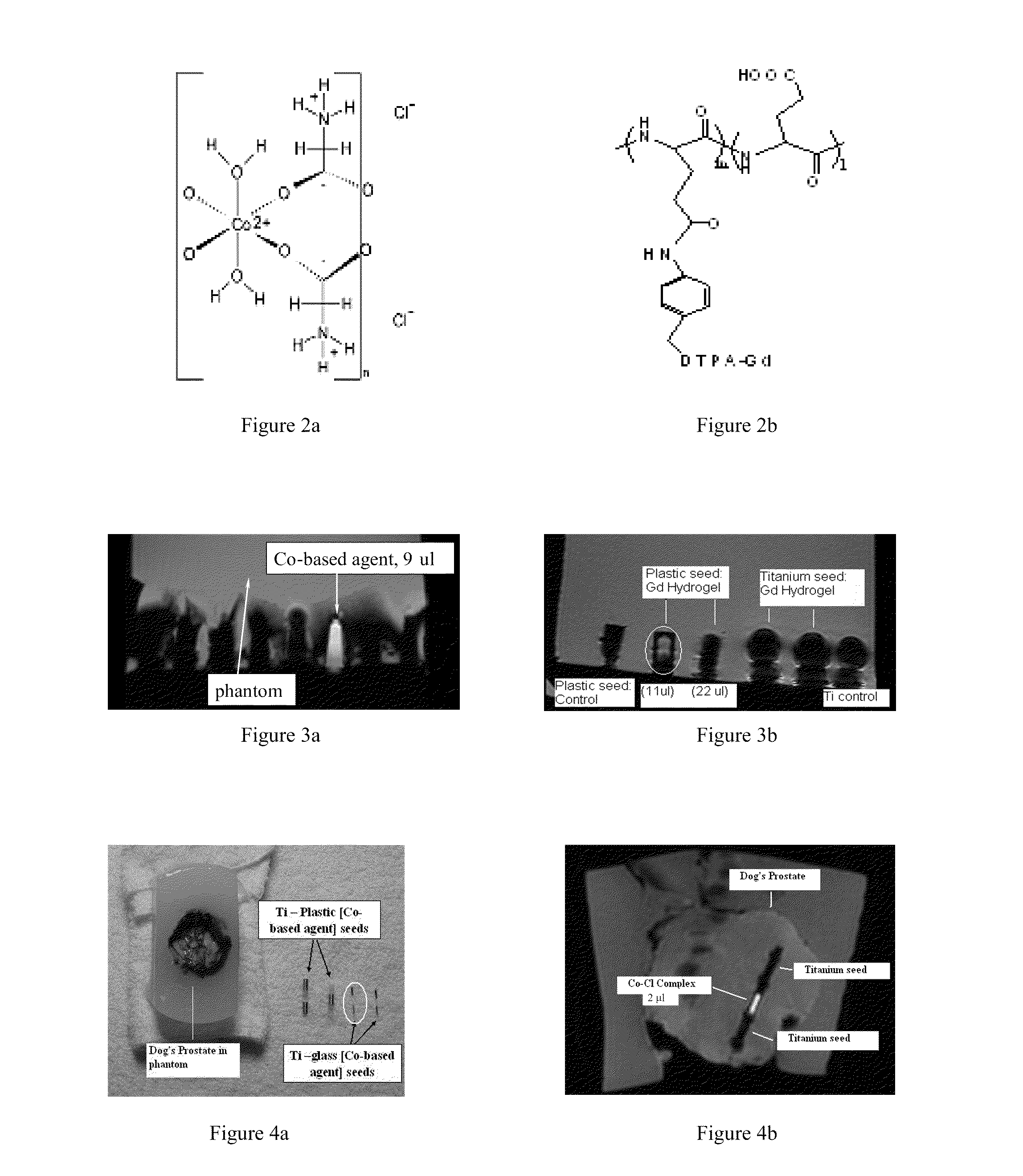
[0198]This example serves to illustrate fabrication of novel MRI visible marker by using biocompatible Poly(methyl methacrylate), (PMMA) and contrast agent C4.

[0199]The C4 water solution with concentration of 0.75% was filtrated through a micro-membrane filter to remove any impurities and injected into the PMMA capillary closed with one side. The dimensions of capillary are: outside diameter, (OD)=0.8 mm, inside diameter, (ID)=0.3 mm and length, (1)=3 mm. The PMMA tap was fastened to the capillary end and locally heated by laser beam to secure the conjunction and to prevent leakage of the contrast agent. Fabricated marker was placed inside the agarose phantom and tested by clinical 1.5 T MRI. The result shows excellent positive MRI signal (see FIG. 34).

example 2

[0200]This example serves to illustrate fabrication of novel MRI visible marker by using biocompatible Poly(methyl methacrylate), (PMMA) and contrast agent C4.

[0201]The C4 water solution with concentration of 0.3% was filtrated through a micro-membrane filter to remove any impurities and injected into the PMMA capillary closed with one side. The dimensions of capillary are: outside diameter, (OD)=0.8 mm, inside diameter, (ID)=0.6 mm and length, (1)=4 mm. The PMMA tap was fastened to the capillary end and locally heated by laser beam to secure the conjunction and to prevent leakage of the contrast agent. Fabricated marker was placed inside the agarose phantom and tested by clinical 1.5 T MRI. The result shows excellent positive MRI signal.

example 3

[0202]This example serves to illustrate fabrication of novel MRI visible marker by using biocompatible Polyetheretherketon, (PEEK) and contrast agent C4.

[0203]The C4 water solution with concentration of 0.75% was filtrated through a micro-membrane filter to remove any impurities and injected into the PEEK capillary closed with one side. The dimensions of capillary are: outside diameter, (OD)=0.8 mm, inside diameter, (ID)=0.6 mm and length, (1)=3 mm. The PEEK tap was fastened to the capillary end and locally heated by laser beam to secure the conjunction and to prevent leakage of the contrast agent. Fabricated marker was placed inside the agarose phantom and tested by clinical 1.5 T MRI. The result shows good positive MRI signal.


PUM

PropertyMeasurementUnit
Concentrationaaaaaaaaaa
Radioactivityaaaaaaaaaa
Relaxation timeaaaaaaaaaa

Abstract

A contrast marker having a casing and a novel MRI contrast agent comprising metal complexes disposed around, within, or abuting the casing is provided. Such contrast markers may be placed in a strand, with or without a therapy seed, to produce a seeded strand useful for imaging and in connection with brachytherapy.

Description

CROSS-REFERENCE TO RELATED APPLICATIONS[0001]This application claims priority to U.S. Pat. App. Ser. No. 60 / 949,157 filed Jul. 11, 2007. This application is incorporated by reference herein it its entirety.FIELD OF INVENTION[0002]The present invention relates generally to the magnetic resonance imaging (MRI) and more specifically, contrast agents and markers and methods of the same.STATEMENT REGARDING FEDERALLY SPONSORED RESEARCH OR DEVELOPMENT[0003]None.THE NAMES OF THE PARTIES TO A JOINT RESEARCH AGREEMENT[0004]None.REFERENCE TO SEQUENCE LISTING[0005]None.BACKGROUND OF THE INVENTION[0006]Magnetic resonance imaging (“MRI”) is the optimal imaging modality for the prostate and surrounding critical organ structures. However, with MRI, the standard titanium radioactive seeds, strands of seeds, and needle tracks appear as black holes (negative contrast) and cannot be accurately localized within the prostate and periprostatic tissue. Without adequate localization of the radioactive seeds...

Claims


Application Information

Patent Timeline
IPC IPC(8): A61K51/12A61K49/08A61P43/00
CPCA61B2019/5454A61N5/1027A61N2005/1023A61B6/508C07F15/065A61K49/18A61K51/1282A61K51/00A61B2090/3954A61P11/00A61P13/08A61P25/00A61P35/00A61P43/00
Inventor FRANK, STEVEN J.MARTIROSYAN, KAREN
Owner BOARD OF RGT THE UNIV OF TEXAS SYST
Why Eureka
Try Eureka