System and method for radiation dose reduction in molecular breast imaging

a molecular breast and radiation dose reduction technology, applied in the field of molecular breast imaging, can solve the problems of reducing the sensitivity of mammography with increasing mammographic density, being criticized as a less than ideal screening method, and being particularly sensitive to limitations of mammography, so as to reduce radiation exposure, reduce noise, and increase radiation exposure

Active Publication Date: 2011-08-25
MAYO FOUND FOR MEDICAL EDUCATION & RES
View PDF2 Cites 9 Cited by
  • Summary
  • Abstract
  • Description
  • Claims
  • Application Information

AI Technical Summary

Benefits of technology

[0007]The present invention overcomes the aforementioned drawbacks by providing a system and method for creating molecular breast images using a reduced radiation exposure, yet yielding images having contrast and noise ratios comparable to molecular breast images created using significantly higher radiation exposures. In particular, a system and method is providing for selectively combining the images associated with each of the detector arrays of an MBI system to produce a composite molecular breast image reduced noise and preserved contrast.
[0008]In accordance with one aspect of the present invention, a molecular breast imaging system is disclosed that includes a first planar gamma detector positioned opposite a second planar gamma detector, each configured to receive photons emitted from a radionuclide present in a subject's breast that is positioned between the first and second gamma detectors. The molecular breast imaging system also includes a processor configured to produce a first detector image associated with photons received by the first gamma detector and a second detector image associated with photons received by the second gamma detector and apply a filter configured to selectively combine the first and second detector images to produce a composite molecular breast image having reduced noise compared to the first and second detector images. The filter is further configured to reduce noise in non-target regions and preserve contrast in target regions.

Problems solved by technology

Despite the success of screening mammography, it is also recognized as a less than ideal screening method.
The limitations of mammography are particularly evident in women with mammographically dense breasts.
The reduced sensitivity of mammography with increasing mammographic density is compounded by the fact that increased breast density is a significant risk factor for breast cancer.
Though MBI has been shown to have a high sensitivity to small lesions, its application in routine breast cancer screen is limited by the radiation dose associated the radiotracer agent.
A variety of radiotracers may be used for MBI, but all of these radiopharmaceuticals deliver a radiation burden that is an order of magnitude larger than that delivered to a patient from a screening mammograph.
Further, radiation dose reduction by simply reducing the amount of radiopharmaceutical administered to the subject can lead to reduced image quality, since fewer photons strike the gamma cameras.

Method used


Image

  • System and method for radiation dose reduction in molecular breast imaging
  • System and method for radiation dose reduction in molecular breast imaging
  • System and method for radiation dose reduction in molecular breast imaging

Examples


Embodiment Construction

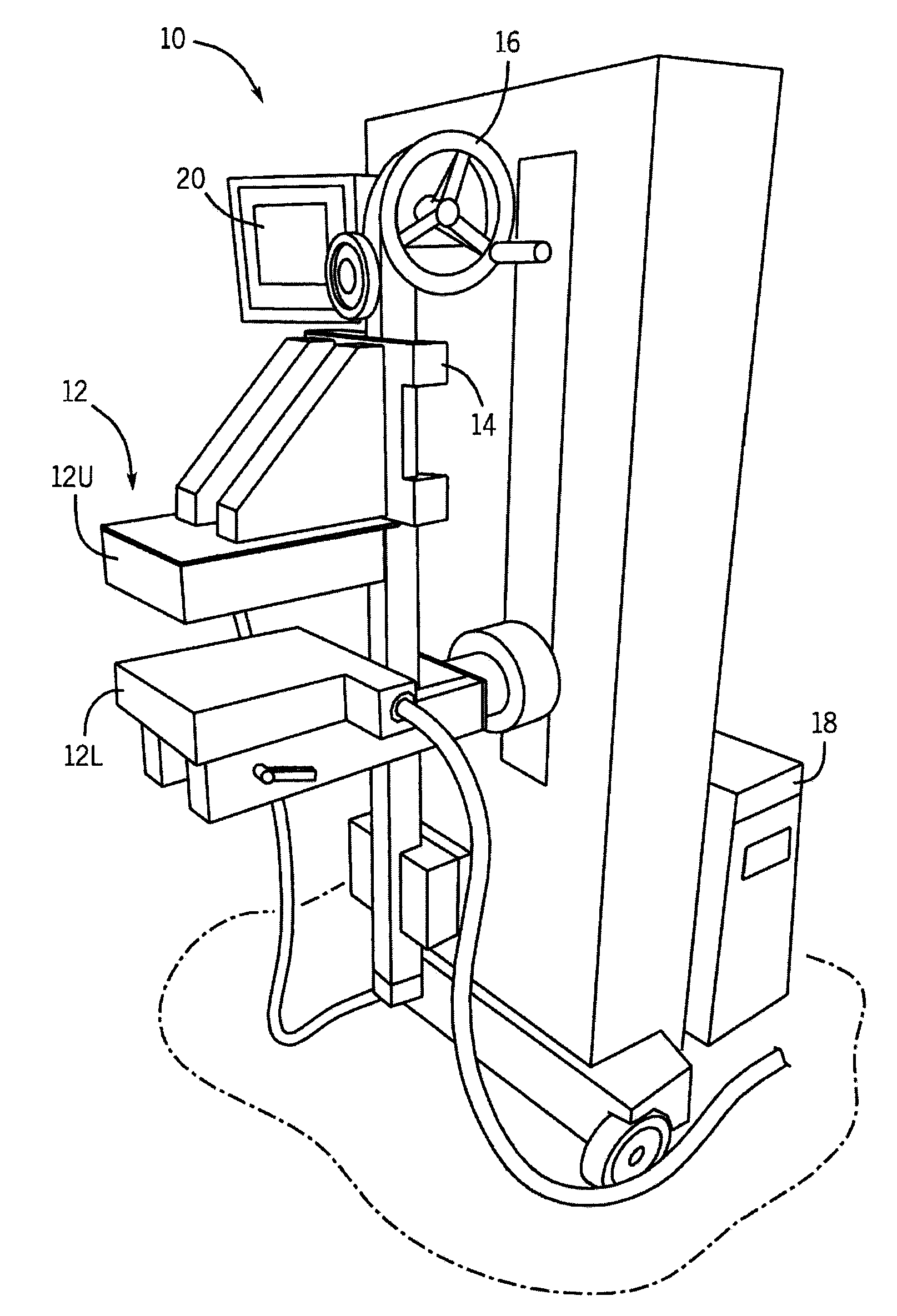
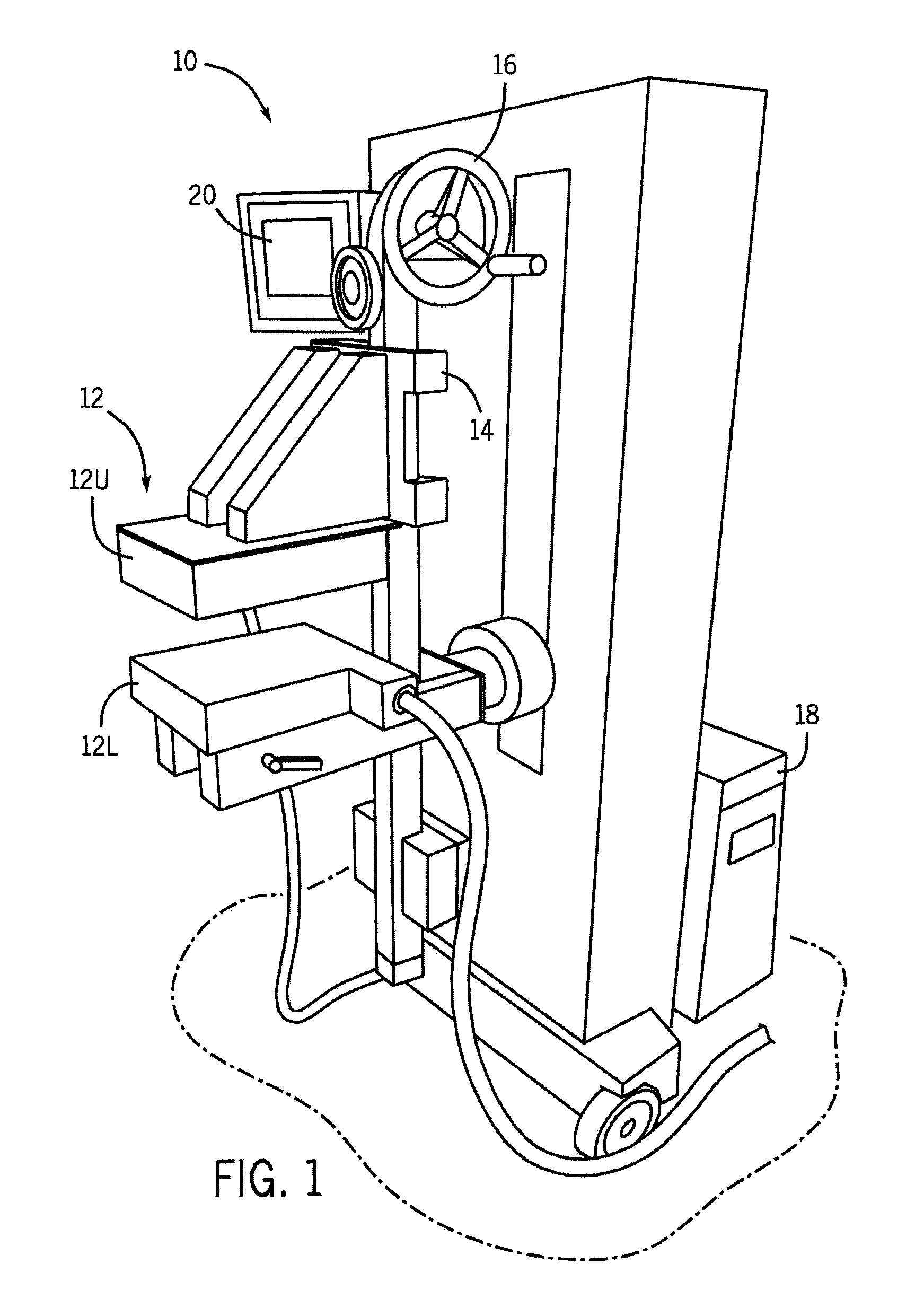
[0016]Referring to FIG. 1, a molecular breast imaging system 10 includes two opposing detectors 12, for example, two cadmium zinc telluride (CZT) detectors (detector heads). In particular, the detector heads 12 include an upper detector head 12U and a lower detector head 12L. Each detector head 12U, 12L is, for example, 20 cm by 16 cm in size and mounted on a modified upright type mammographic gantry 14.

[0017]The relative position of the detector heads 12 can be adjusted using a user control 16. Specifically, the detector head assemblies 12 are, preferably, designed to serve as a compression mechanism. Accordingly, this system configuration reduces the maximum distance between any lesion in the breast and either detector head 12 to one-half of the total breast thickness, potentially increasing detection of small lesions without additional imaging time or dose. The MBI system 10 includes a processor 18 for processing the signals acquired by the detector heads 12 to produce an image, ...


PUM


Abstract

A system and method is provided for analyzing detector data acquired during molecular breast imaging (MBI) that reduces a patient's radiation does without a reduction in image quality. The method processes and combines initial images acquired by the two gamma-camera detectors of the MBI system to produce a hybrid or composite image set having reduced noise and improved contrast. The composite image provides image quality comparable to that of traditional MBI methods, but at a lower patient radiation dose.

Description

CROSS-REFERENCE TO RELATED APPLICATIONS[0001]This application is based on, claims the benefit of, and incorporates herein by reference U.S. Provisional Application Ser. No. 61 / 110,639, filed Nov. 3, 2009, and entitled “SYSTEM AND METHOD FOR RADIATION DOSE REDUCTION IN MOLECULAR BREAST IMAGING.”FIELD OF THE INVENTION[0002]The present invention relates to systems and methods for molecular breast imaging and, more particularly, to a system and method for reducing the dose of radioactivity to which a patient is subjected while preserving image quality.BACKGROUND OF THE INVENTION[0003]Breast cancer is one of the most common forms of cancer and a leading cause of cancer death. Breast cancer screening has been recommended for many decades and several large scale studies have demonstrated a clear benefit to screening, particularly for women over the age of fifty. The combination of early detection and improved therapy has resulted in a significant reduction in breast cancer mortality.[0004]...

Claims


Application Information

Patent Timeline
Patent Type & Authority Applications(United States)
IPC IPC(8): A61B6/00G06K9/00
CPCA61B6/4258G01T1/00
Inventor O'CONNOR, MICHAEL K.HRUSKA, CARRIE B.
Owner MAYO FOUND FOR MEDICAL EDUCATION & RES
Why Eureka
Try Eureka