
Shape Modeling and Detection of Catheter

a catheter and shape technology, applied in the field of fluoroscopic imaging, can solve the problems of difficult catheter detection in such fluoroscopic images, and achieve the effect of reducing parameters associated

Active Publication Date: 2011-11-24
SIEMENS HEALTHCARE GMBH
View PDF5 Cites 2 Cited by
  • Summary
  • Abstract
  • Description
  • Claims
  • Application Information

AI Technical Summary

Benefits of technology

[0007]In an embodiment of the present invention, the trained shape model is fitted in a learned subspace detected using principle component analysis to reduce parameters associated with the trained shape models.

Problems solved by technology

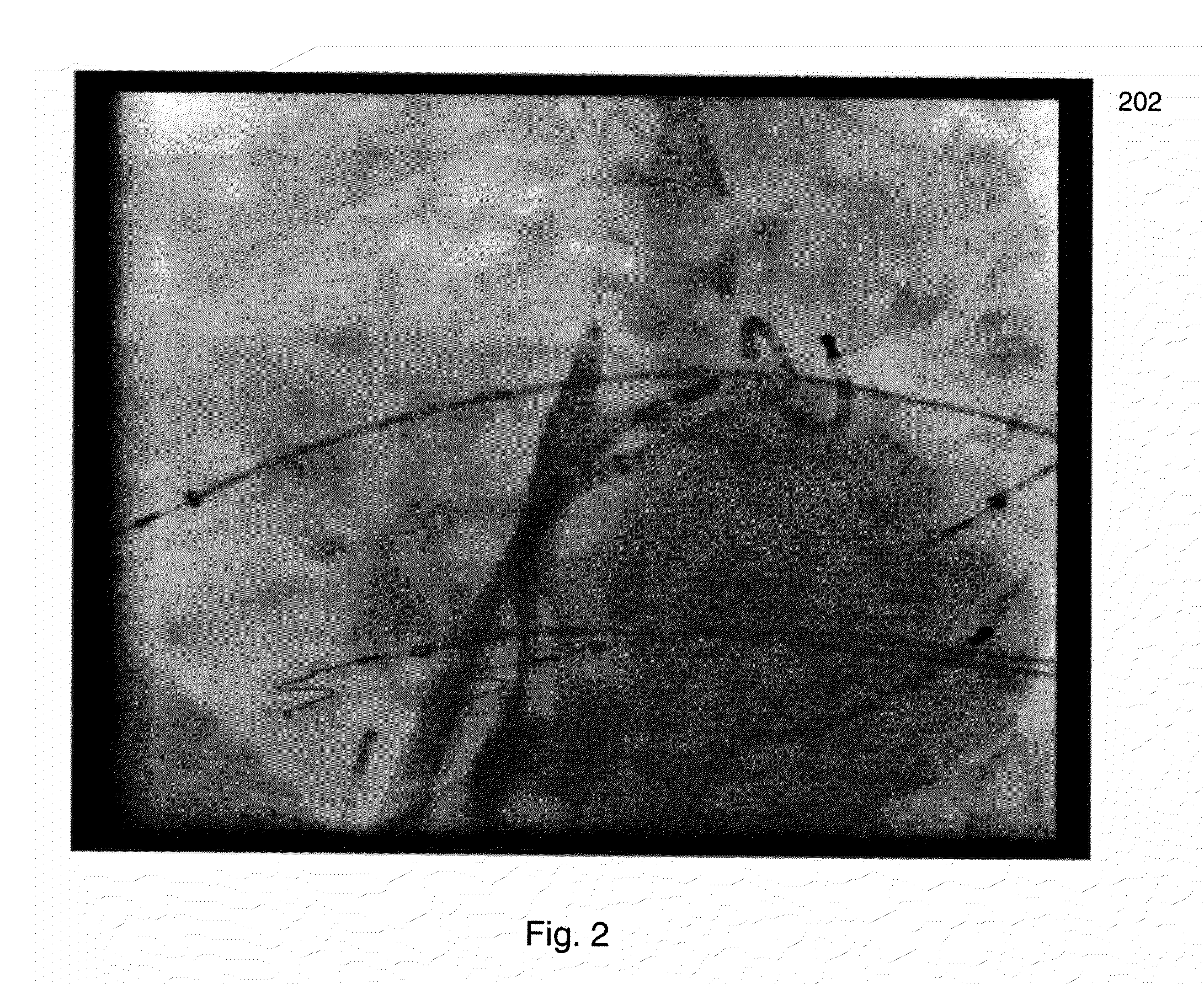
The detection of catheters in such fluoroscopic images is difficult due to varying imaging quality of the fluoroscopic images.

Method used


Image

  • Shape Modeling and Detection of Catheter
  • Shape Modeling and Detection of Catheter
  • Shape Modeling and Detection of Catheter

Examples


Embodiment Construction

[0019]The present invention is directed to a method and system for detecting and modeling a catheter in a fluoroscopic image. Embodiments of the present invention are described herein to provide a visual understanding of the catheter detection and modeling method. A digital image often includes digital representations of objects or shapes. The digital representation of an object is often described herein in terms of identifying and manipulating the objects. Such manipulations are virtual and carried out in the memory or other circuitry / hardware of a computer system. Accordingly, it is to be understood that embodiments of the present invention may be performed within a computer system using data stored within the computer system.

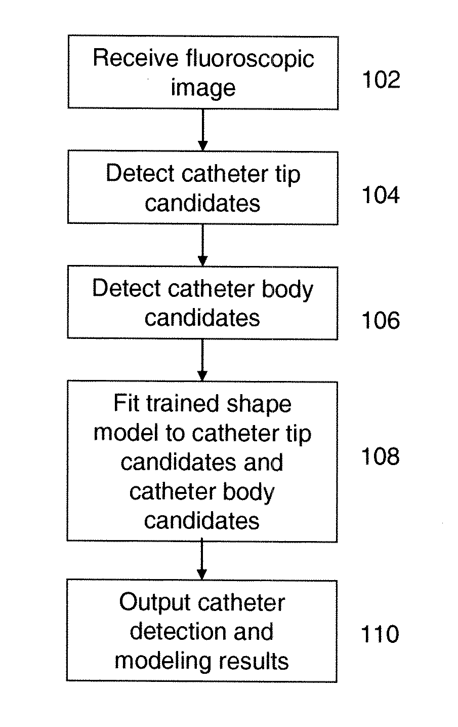
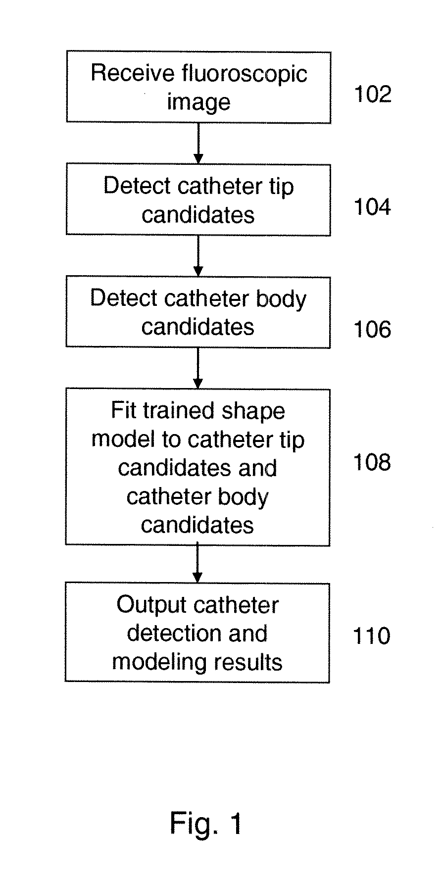
[0020]FIG. 1 illustrates a method for detecting and modeling a catheter in a fluoroscopic image according to an embodiment of the present invention. At step 102, a fluoroscopic image is received. The fluoroscopic image may be a part of a fluoroscopic image se...


PUM


Abstract

A method and system for detecting and modeling a catheter in a fluoroscopic image is disclosed. Catheter tip candidates and catheter body candidates are detected in the fluoroscopic image. One of a plurality of trained shape models is fitted to the catheter tip candidates and the catheter body candidates in order to model a shape of the catheter in the fluoroscopic image.

Description

[0001]This application claims the benefit of U.S. Provisional Application No. 61 / 346,196, filed May 19, 2010, the disclosure of which is incorporated herein by reference.BACKGROUND OF THE INVENTION[0002]The present invention relates to fluoroscopic imaging, and more particularly, to the detection and modeling of a catheter within fluoroscopic image sequences.[0003]The detection of catheters is important in a variety of medical applications. Catheters are tubes that may be inserted into a body cavity, duct, or vessel. Some types of catheter may be used to generate an electrical map of a heart to assist medical professionals in identifying locations of abnormal electrical activity within the heart. These types of catheters may be inserted into arteries and guided to the heart. Medical procedures using catheters are typically monitored using fluoroscopic imaging, in which a sequence of fluoroscopic images is acquired in real-time during the procedure. The detection of catheters in such...

Claims


Application Information

Patent Timeline
Patent Type & Authority Applications(United States)
IPC IPC(8): A61B5/05
CPCA61B6/12A61B6/5211A61B6/487G16H50/20
Inventor YI, SHENGCHEN, TERRENCEWANG, PENGCOMANICIU, DORIN
Owner SIEMENS HEALTHCARE GMBH
Learn More