Methods and apparatus for localized administration of inhibitory moieties to a patient

a technology of inhibitory moieties and localization, applied in radiation therapy, pharmaceutical non-active ingredients, therapy, etc., can solve the problems of life-threatening complications, physiologic state, and organ fed by that artery suffers ischemia, and achieve the effect of inhibiting unwanted cellular growth

Inactive Publication Date: 2011-12-29
ABBOTT LAB VASCULAR ENTERPRISE
View PDF0 Cites 2 Cited by
  • Summary
  • Abstract
  • Description
  • Claims
  • Application Information

AI Technical Summary

Benefits of technology

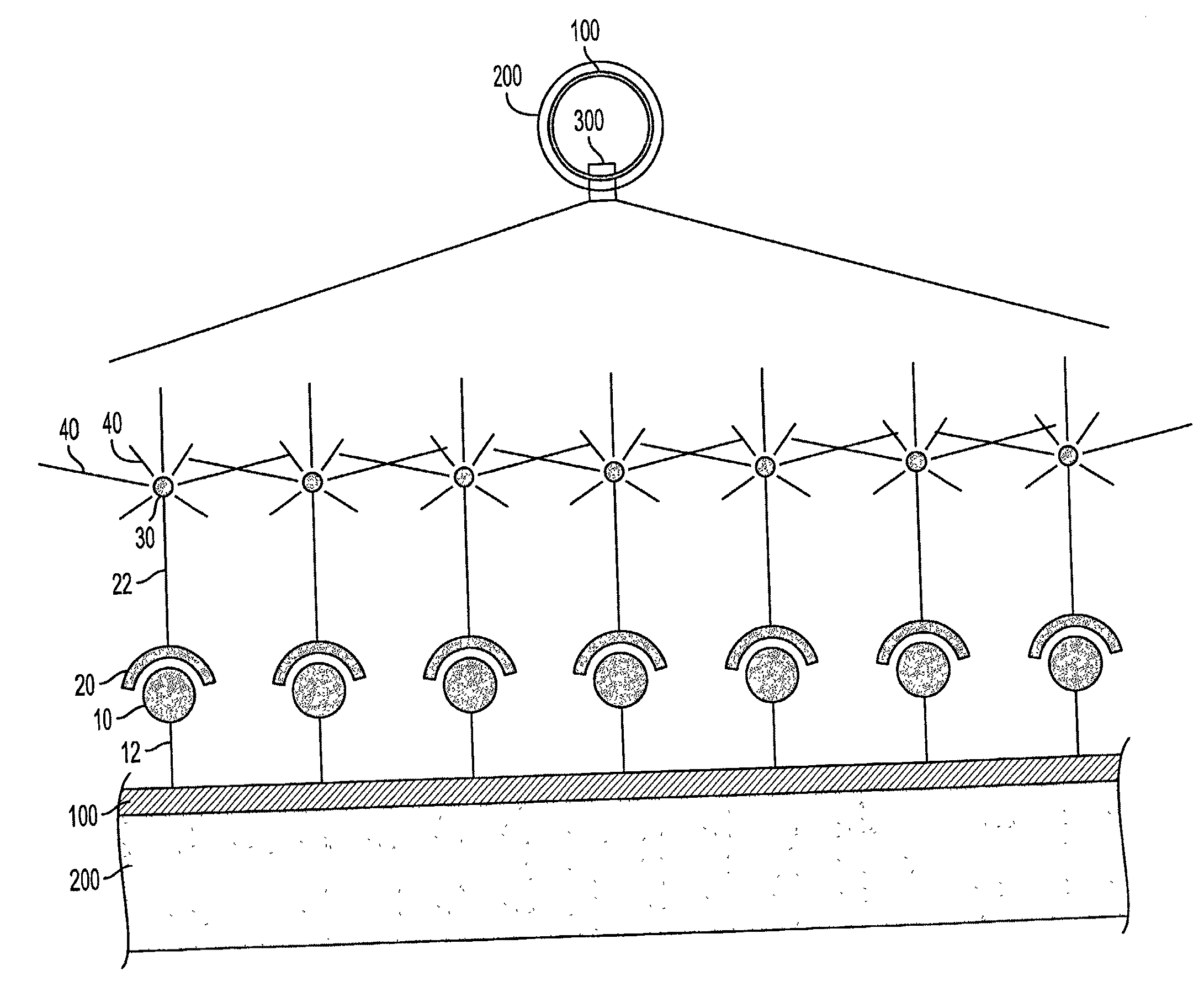
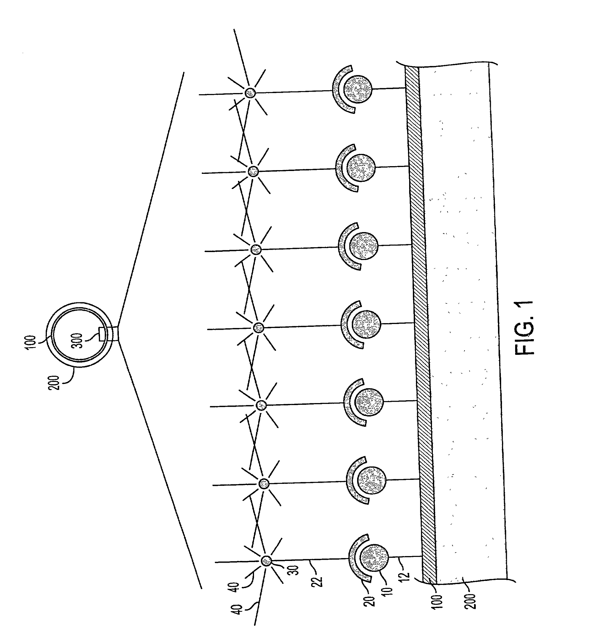
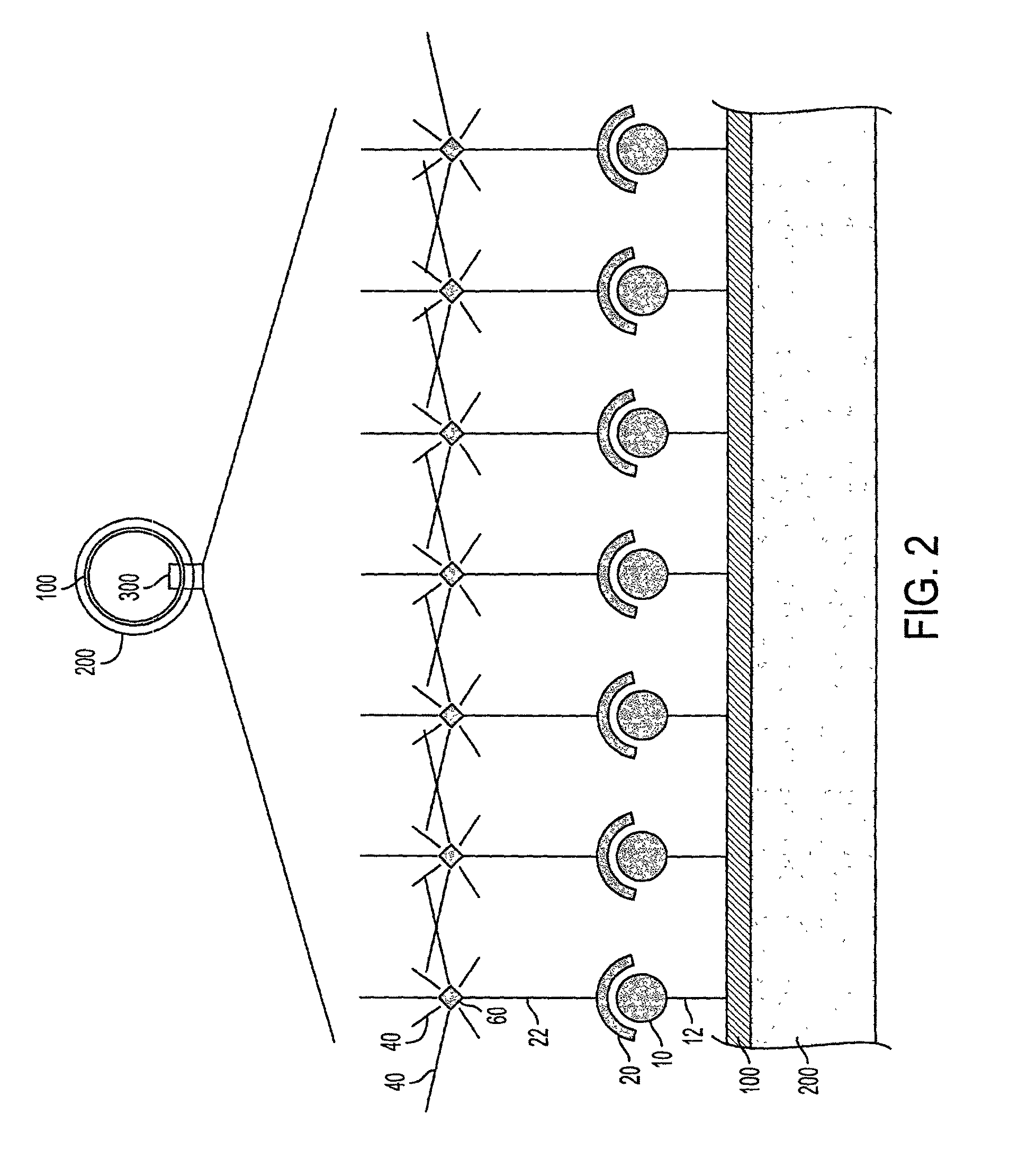
[0041]It is, therefore, a further object of the present invention to provide methods of localized administration to a patient of such radioactive and other moieties for the purpose of rendering an implanted medical device capable of inhibiting unwanted cellular growth.
[0042]It is yet a further object of the present invention to provide medical devices that can be rendered capable of inhibiting unwanted cellular growth after implantation in a patient by the methods of localized administration provided herein.
[0044]It is a further object of the present invention to provide stents that can be rendered capable of inhibiting restenosis after implantation in a patient by the methods of localized administration of radioactive moieties and other moieties capable of inhibiting restenosis.

Problems solved by technology

Continued growth and development of a plaque can result in life threatening complications.
As a result, the organ fed by that artery suffers ischemia, a physiologic state characterized by inadequate oxygen and nutrient supplies to meet the metabolic need, as well as diminished removal of metabolic waste products.
At minimum, ischemia compromises the function of the organ, and if severe enough, can result in tissue and organ death.
Stenosis of the coronary arteries results in ischemia of the heart tissue which can precipitate myocardial infarction (heart attack), the death of those parts of the heart muscle dependent on the particular coronary artery that is blocked.
Particularly dangerous for the patient with atherosclerosis of the coronary arteries is the occurrence of an acute plaque change.
In both cases the blood is exposed to highly thrombogenic substances in or near the plaque, causing platelet adhesion, aggregation, activation and release of molecules that further promote aggregation, resulting a rapid feed-forward cycle causing a clot to form over the disrupted plaque.
At best, healing of the thrombus and disrupted plaque results in growth of the overall plaque lesion; at worst, the thrombus completely occludes the artery.
In the coronary artery this often results in myocardial infarction, but can also cause sudden cardiac death if the ischemia triggers ventricular fibrillation.
The thrombus can also embolize, throwing off small fragments that are carried downstream in the blood to lodge in smaller arteries, which can also result in heart attack and stroke.
In the first years PTCA was practiced, acute restenosis, an unwelcome complication of PTA, arose in some patients.
This life threatening complication required repetition of the PTCA procedure or resort to the traditional coronary artery bypass graft procedure (CABG) to regain perfusion.
Although evidence suggests that catheter-based brachytherapy may be effective, this approach presents problems.
One limitation involves the tradeoff between the time necessary for the optimal dose to be absorbed and the degree of radioactivity of the source.
However, the lower the activity of the source, the greater the amount of time the catheter must be in-place in the patient for the required dose of radiation to be absorbed.
Increased dwell-time reduces the total number of patients that a busy angioplasty team can treat, but more importantly, increases the opportunity for unwanted side effects, such as thrombosis formation, to occur.
Thus, using the catheter-based technique, there is no ability to administer the same dose over substantially longer periods.
A common limitation of all these approaches whereby the stent is made radioactive first and subsequently implanted into a patient is that it is not generally possible to prepare the radioactive stent for implantation in the hospital.
This raises logistical and safety problems associated with shielding the radioactive stent assembly during distribution to the end user.
Logistical problems are exacerbated when the radioactive stent comprises a short-half life radionuclide, such as 32P (t1 / 2=14.3 days), because reliable availability of a stent with a predefined radiation dose would require a well coordinated manufacturing and distribution system to ensure that the stent reaches the end user before the radioactivity decays to ineffective levels.
This would make radioactive stents much more expensive to the end user.
At minimum, this results in unnecessary full-body exposure of the patient to radioactivity.
Also, as a consequence of systemic administration, the radioactive substance becomes highly diluted in the volume of circulating blood.
Further, the circulating concentration does not remain constant, but declines as the substance is metabolized, principally by the kidneys and liver.
In the second instance, there exists the risk of potentially toxic effects from introducing a large amount of a foreign substance into the body.
Administration of a larger amount of the radioactive substance, of course, also increases the whole body exposure.
However, this has the consequence of reducing the efficacy of the stent once it has become radioactive.
Systemic administration of large quantities of a radioactive substance also suffers from the problem that, as the substance is metabolized and excreted, the urine and feces become contaminated with radioactivity, necessitating expensive and inconvenient collection and disposal.

Method used


Image

  • Methods and apparatus for localized administration of inhibitory moieties to a patient
  • Methods and apparatus for localized administration of inhibitory moieties to a patient
  • Methods and apparatus for localized administration of inhibitory moieties to a patient

Examples


example 1

Preparing a Biotin Labeled Stent

[0340]Stainless steel coronary artery stents, such as the JOSTENT® Flex stent, in their unexpanded state are prepared for biotin immobilization by first subjecting the stents to electrolysis in 5×SSC solution to form an oxide layer. Alternatively, an oxide layer is formed by immersion of the stents into a perchloric acid bath. After oxidation, the stents are rinsed in 100% ethanol and air dried in a 50° C. oven.

[0341]Thereafter, the oxidized stents are reacted with 10% 3-aminopropyltriethoxysilane (APS) in toluene. The stents are placed into a glass reaction dish, after which toluene is added to completely cover the stents, and heated to 50-60° C. on a heat plate. APS is added to a final concentration of 10%. The solution is gently mixed and allowed to continue for 30minutes. The solution is removed, and the stents are rinsed three times with large volumes of toluene, followed by rinsing three times in large volumes of 100% ethanol, followed by air dr...


PUM


Abstract

Methods and devices are provided for the localized administration to a patient of moieties effective for inhibiting unwanted cellular growth, including restenosis of an artery treated with a stent implant for blockage of blood flow by an atherosclerotic lesion. After implantation of a medical device capable of moiety-binding, moieties effective at inhibiting unwanted cellular growth are administered locally to a patient. In this manner the deleterious side-effects of systemic administration of moieties are avoided. Upon localized administration, the moieties bind the medical device, rendering the medical device itself capable of inhibiting unwanted cellular growth. According to an embodiment, after implantation of a stent, radioactive moieties specific for receptors immobilized on the stent surface are locally administered using a balloon perfusion catheter. The moieties bind specifically the receptors, becoming immobilized thereto, thereby rendering the stent radioactive and effective for inhibiting restenosis.

Description

CROSS-REFERENCE TO RELATED APPLICATIONS[0001]This application claims priority from the copending U.S. patent application Ser. No. 09 / 303,849, filed May 3, 1999, and Ser. No. 09 / 442,591, filed Nov. 17, 1999. This application also claims priority from the German patent applications No. 19819635.0, filed May 5, 1998, and No. 19853067.6, filed Nov. 17, 1998. The disclosure of each of the afore-referenced priority patent applications is incorporated by reference herein in its entirety.FIELD OF THE INVENTION[0002]The present invention relates generally to the inhibition of unwanted cellular growth in the vicinity of medical devices implanted in a patient. More particularly, the present invention relates to medical devices capable of being made radioactive, or otherwise capable of inhibiting unwanted cellular growth, and methods of making such medical devices radioactive or otherwise growth inhibitory. Yet more particularly, the present invention relates to stents capable of being made rad...

Claims


Application Information

Patent Timeline
Patent Type & Authority Applications(United States)
IPC IPC(8): A61F2/06A61F2/82
CPCA61F2/82A61F2210/0095A61K47/48992A61N5/1002A61L31/18A61L2300/416A61L2300/44A61L31/16A61K47/6957
Inventor OEPEN, RANDOLF VON
Owner ABBOTT LAB VASCULAR ENTERPRISE
Why Eureka
Try Eureka