Method and System for Tracking of a Virtual Electrode on a Coronary Sinus Catheter in Fluoroscopic Images

a virtual electrode and coronary sinus technology, applied in the field of detection and tracking of coronary sinus catheters in fluoroscopic images, can solve the problems of large displacement, cluttered background, and difficulty in conventional tracking algorithms in the presence of large image variations, and achieve the effect of robust and fast methods

Inactive Publication Date: 2012-09-13
SIEMENS HEATHCARE GMBH
View PDF11 Cites 15 Cited by
  • Summary
  • Abstract
  • Description
  • Claims
  • Application Information

AI Technical Summary

Benefits of technology

[0005]Embodiments of the present invention provide a robust and fast method to track a virtual electrode on a coronary sinus (CS) catheter that is more proximal than the most proximal electrode in a sequence of fluoroscopic images. E

Problems solved by technology

However, the heart is not a static structure, and the relative motion of mapping and reference catheters can lead to significant displacements.
However,

Method used


Image

  • Method and System for Tracking of a Virtual Electrode on a Coronary Sinus Catheter in Fluoroscopic Images
  • Method and System for Tracking of a Virtual Electrode on a Coronary Sinus Catheter in Fluoroscopic Images
  • Method and System for Tracking of a Virtual Electrode on a Coronary Sinus Catheter in Fluoroscopic Images

Examples


Embodiment Construction

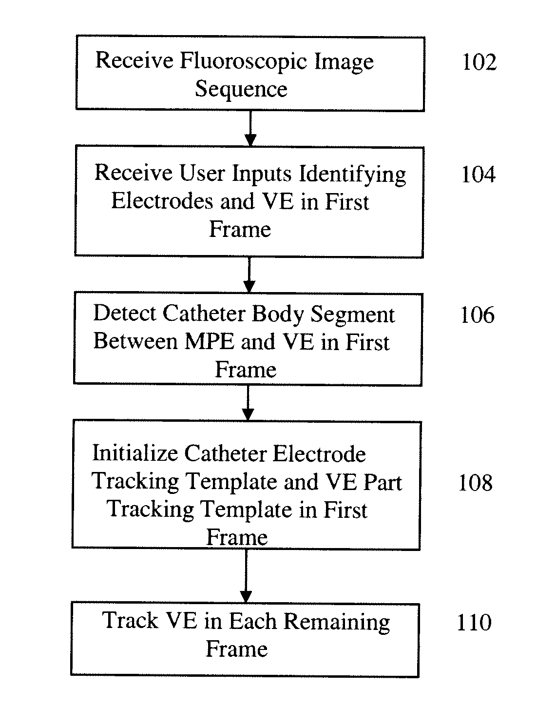
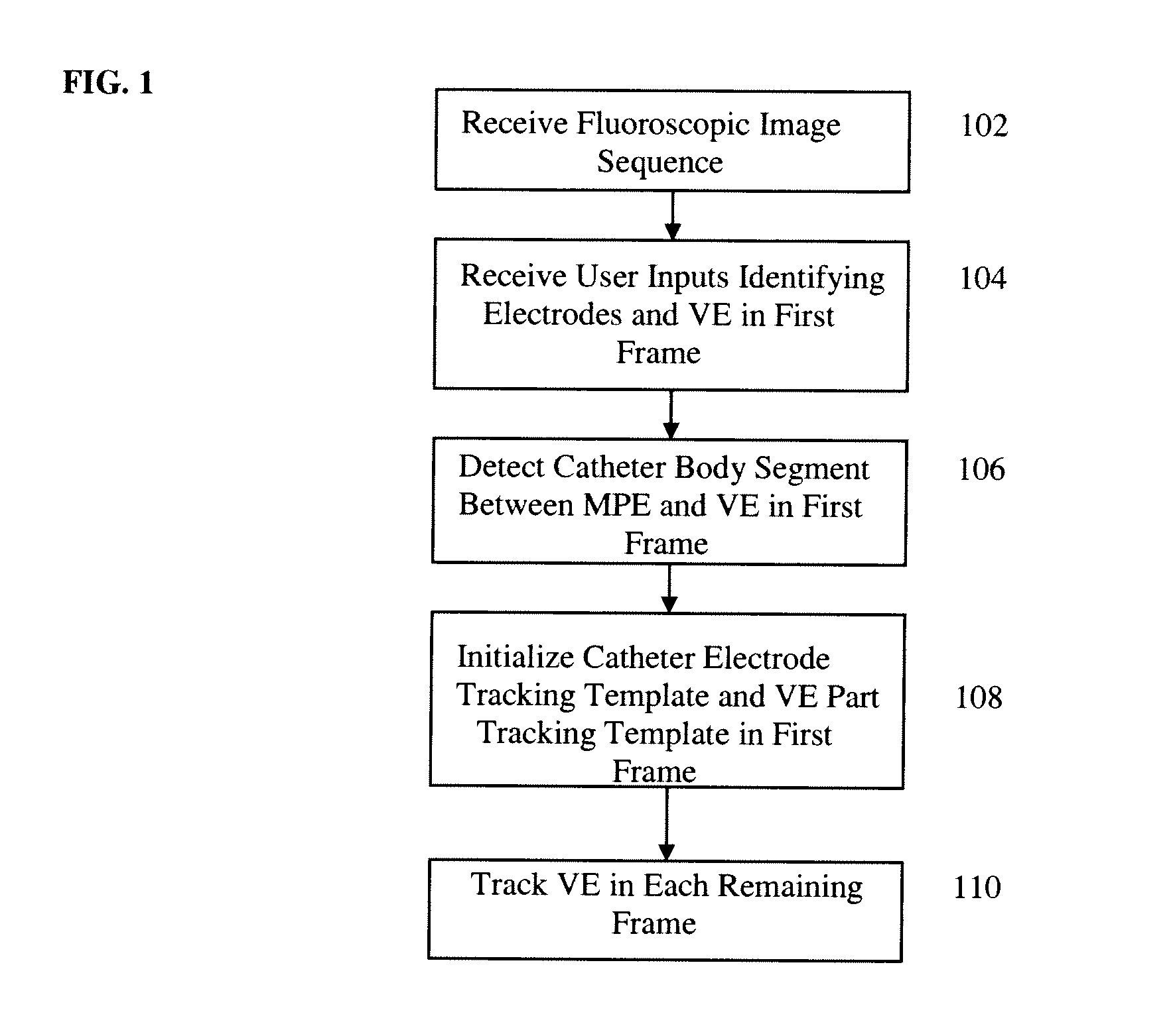
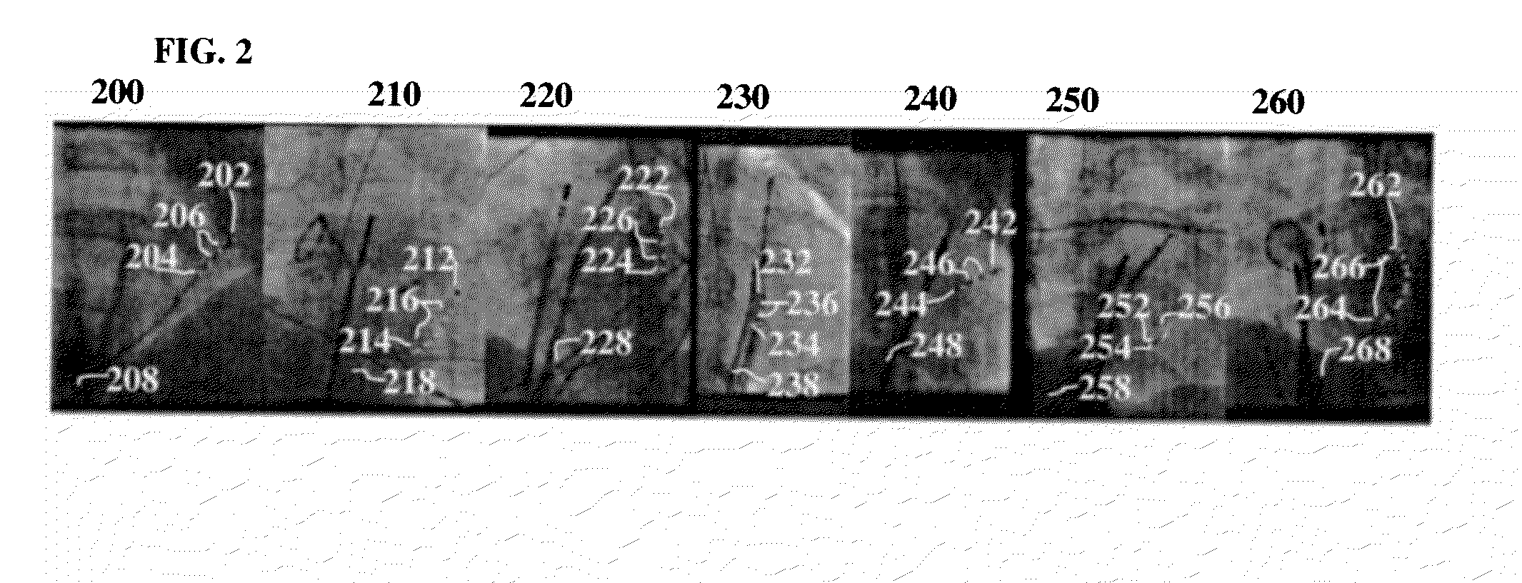
[0016]The present invention relates to a method and system for tracking a virtual electrode on a coronary sinus (CS) catheter in fluoroscopic images. Embodiments of the present invention are described herein to give a visual understanding of the CS catheter virtual electrode tracking method. A digital image is often composed of digital representations of one or more objects (or shapes). The digital representation of an object is often described herein in terms of identifying and manipulating the object. Such manipulations are virtual manipulations accomplished in the memory or other circuitry / hardware of a computer system. Accordingly, is to be understood that embodiments of the present invention may be performed within a computer system using data stored within the computer system.

[0017]Tracking the coronary sinus (CS) catheter can help compensate respiratory and cardiac motion for 3D overlay to assist in positioning an ablation catheter in atrial fibrillation (AF) treatments. Howe...


PUM


Abstract

A method and system for detecting a virtual electrode (VE) on a coronary sinus (CS) catheter in a fluoroscopic image sequence is disclosed. User inputs indicating locations of CS catheter electrodes and a location of a VE are received. A catheter electrode model and a VE part model is initialized in a first frame of the fluoroscopic image sequence. The VE is tracked by detecting electrode position candidates and catheter body point candidates in the subsequent frames of the fluoroscopic image sequence using respective trained detectors, tracking the catheter electrode model in the subsequent frames based on the detected electrode position candidates, generating VE part hypotheses in the subsequent frames based on detection of the most proximal electrode (MPE) in each subsequent frame, calculating a probability score for each of the VE part hypotheses, and selecting an VE part hypothesis with the highest probability score.

Description

[0001]This application claims the benefit of U.S. Provisional Application No. 61 / 449,862, filed Mar. 7, 2011, the disclosure of which is herein incorporated by reference.BACKGROUND OF THE INVENTION[0002]The present invention relates to detection and tracking of a coronary sinus catheter in fluoroscopic images, and more particularly, to detecting and tracking a virtual electrode on a coronary sinus catheter in fluoroscopic images to assist in atrial fibrillation ablation treatment.[0003]Atrial fibrillation (AF) is a rapid, highly irregular heartbeat caused by abnormalities in the electrical signals generated by the atria of the heart. It is the most common cardiac arrhythmia (abnormal heart rhythm) and involves the two upper chambers (atria) of the heart. Surgical and catheter-based therapies have become common AF treatments throughout the world. Catheter ablation modifies the electrical pathways of the heart in order to treat AF. To measure electrical signals in the heart and assist...

Claims


Application Information

Patent Timeline
IPC IPC(8): A61B6/12G06V10/75
CPCA61B6/12A61B6/487G06K9/64G06T7/204G06T2207/30048G06T2207/10121G06T2207/20081G06T2207/30021G06T7/208G06T7/248G06T7/277G06V10/75
Inventor WU, WENCHEN, TERRENCESTROBEL, NORBERTFUNKA-LEA, GARETHCOMANICIU, DORIN
Owner SIEMENS HEATHCARE GMBH
Why Eureka
Try Eureka