Determining velocity of cerebrospinal fluid by magnetic resonance imaging

a magnetic resonance imaging and cerebrospinal fluid technology, applied in the field of image processing apparatus and magnetic resonance imaging apparatus, can solve the problems of inapplicability, insufficient image quality, and inability to read images in time and effort,

Active Publication Date: 2012-11-29
TOKAI UNIV +1
View PDF8 Cites 5 Cited by
  • Summary
  • Abstract
  • Description
  • Claims
  • Application Information

AI Technical Summary

Problems solved by technology

However, when observing dynamics of a CSF by using the phase shift method, there are problems as described below.
In such case, to what extent the maximum velocity is, and how far a CSF in a certain portion reaches within a certain time are influential and meaningful, so that only an image of an average velocity in a relatively long time range obtained by the phase shift method is substantially insufficient.
Moreover, according to the phase shift method, fluctuations in the velocity at the time of collecting each echo signal are large, consequently, ghost artifact tends to appear in the phase encoding direction on a reconstructed image, and the image quality has a limitation.
However, the method consumes time and efforts for reading images and is not practical, because images are not provided in a sorted manner for observing dynamics of a CSF, and a number of images have to be referred.
As described above, according to conventional methods, for example, the phase shift method, velocity variations of a body fluid that are not cyclical, such as velocity variations of a CSF, cannot be faithfully imaged.

Method used


Image

  • Determining velocity of cerebrospinal fluid by magnetic resonance imaging
  • Determining velocity of cerebrospinal fluid by magnetic resonance imaging
  • Determining velocity of cerebrospinal fluid by magnetic resonance imaging

Examples


first embodiment

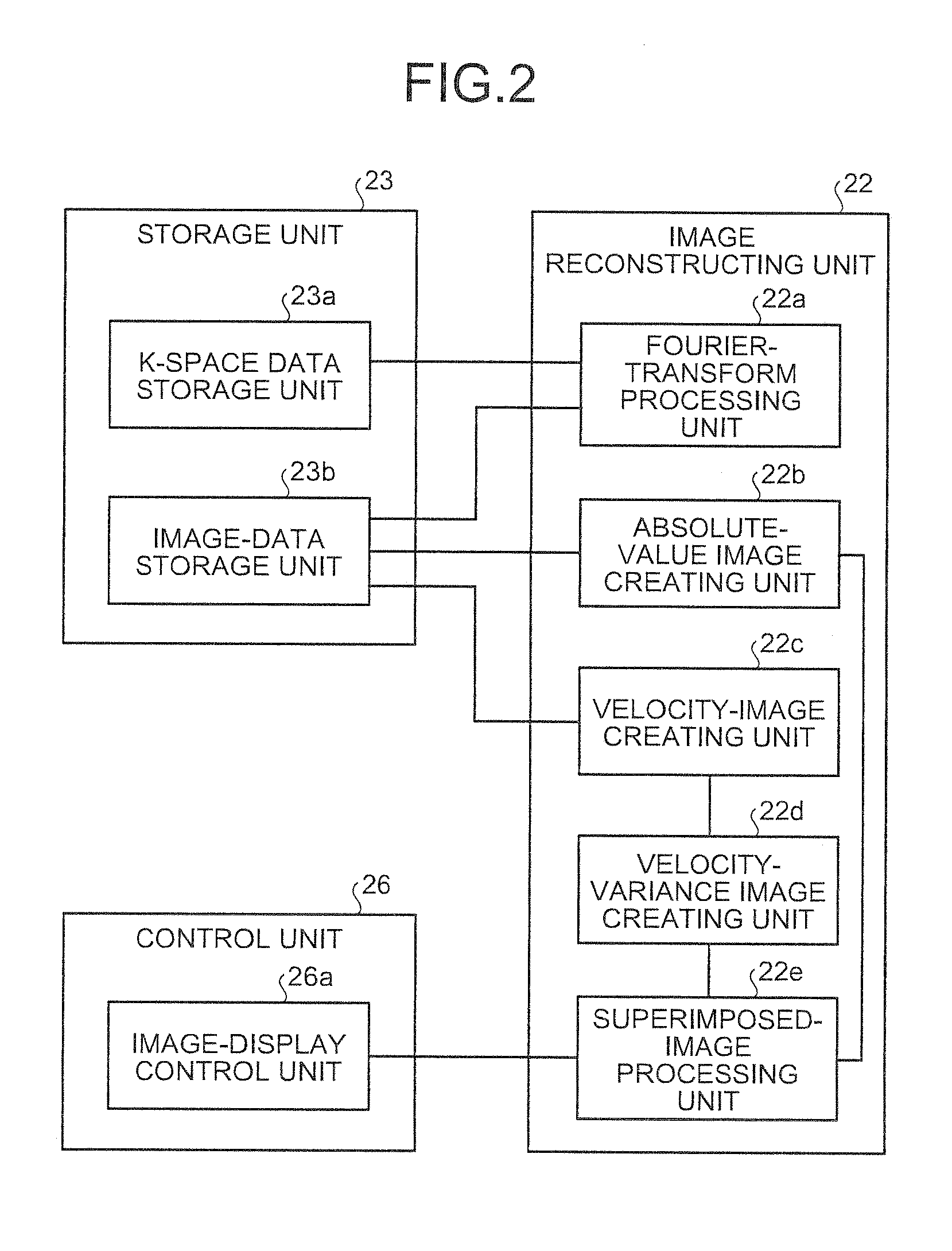
[0059] the Fourier-transform processing unit 22a reconstructs a plurality of images S(ra, i) as shown in FIG. 4 by performing reconstruction processing, such as a discrete two-dimensional Fourier transform, on respective k-space data of echo signals obtained through one shot as explained above. According to FIG. 4, ra denotes a vector that indicates a position on an image, and i denotes the order of collection. According to the images S(ra, i), the phase of a pixel shifts proportionally to the velocity.

[0060]The absolute-value image creating unit 22b creates absolute-value images from images reconstructed by the Fourier-transform processing unit 22a. Specifically, the absolute-value image creating unit 22b creates an absolute-value image I(ra, i) given by Expression (1) described below with respect to each of the images S(ra), i) reconstructed by the Fourier-transform processing unit 22a.

I(ra,i)=abs{S(ra),i)}  (1)

[0061]Furthermore, the absolute-value image creating unit 22b creates...

second embodiment

[0083]FIG. 8 is a functional block diagram of a configuration of the image reconstructing unit, the storage unit, and the control unit according to the For convenience of explanation, functional units that play roles similar to those of the units shown in FIG. 2 are assigned with the same reference numerals, and detailed explanations of them are omitted.

[0084]As shown in FIG. 8, an image reconstructing unit 122 according to the second embodiment particularly includes a Fourier-transform processing unit 122a, an absolute-value image creating unit 122b, a signal-strength variance image creating unit 122d, and a superimposed-image processing unit 122e.

[0085]The Fourier-transform processing unit 122a reconstructs an image by performing reconstruction processing, such as a discrete two-dimensional Fourier transform, on k-space data stored in the k-space data storage unit 23a.

[0086]According to the second embodiment, the Time-SLIP method is used as an imaging method capable of obtainin...


PUM


Abstract

A velocity-image creating unit creates a velocity image that indicates a distribution of velocity components with respect to each of a plurality of images obtained by repeating a plurality of number of times Echo Planar Imaging (EPI) that is capable of obtaining velocity components of a Cerebrospinal Fluid (CSF) flowing inside a subject. A velocity-variance image creating unit calculates variance of velocity components along the time sequence by same position on velocity images by using a plurality of created velocity images. A superimposed-image processing unit then superimposes the distribution of the variance of the velocity components according to the velocity-variance image on an average absolute-value image, and an image display unit displays a superimposed image.

Description

CROSS-REFERENCE TO RELATED APPLICATIONS[0001]This application is a division of U.S. patent application Ser. No. 12 / 572,761 filed Oct. 2, 2009; and this application is based upon and claims the benefit of priority from the prior Japanese Patent Application No. 2008-258963 filed on Oct. 3, 2008, the entire contents of both of which are incorporated herein by reference.BACKGROUND[0002]1. Technical Field[0003]The present exemplary embodiment relates to an image processing apparatus, a magnetic resonance imaging apparatus, and an image processing method for processing an image obtained by imaging an inside of a subject. Particularly, the present exemplary embodiment relates to an image processing apparatus, a magnetic resonance imaging apparatus, and an image processing method according to which a distribution of velocity variations of a body fluid can be faithfully imaged even if the velocity variations of the fluid are not cyclical, such as velocity variations of a Cerebrospinal Fluid ...

Claims


Application Information

Patent Timeline
Patent Type & Authority Applications(United States)
IPC IPC(8): A61B5/055
CPCA61B5/055A61B5/7257G01R33/5602G01R33/5608G01R33/5616G06T2207/30016G01R33/56316G06T5/50G06T7/208G06T2207/10088G06T2207/20221G01R33/5617G06T7/277
Inventor YAMADA, SHINYAKANAZAWA, HITOSHI
Owner TOKAI UNIV
Why Eureka
Try Eureka