Treatment of Urinary Incontinence with Regenerative Glue

a technology of regenerative glue and urinary incontinence, which is applied in the field of treatment of urinary incontinence with regenerative glue, can solve the problems of stress urinary incontinence, which is associated with a disturbing level, and achieves the effect of meliorating one or more symptoms

Inactive Publication Date: 2014-08-14
UNIVERSITY HOSPITALS OF CLEVELAND CLEVELAND
View PDF1 Cites 9 Cited by
  • Summary
  • Abstract
  • Description
  • Claims
  • Application Information

AI Technical Summary

Benefits of technology

The present invention describes how much of these substances are needed to treat diseases or conditions like infections. This helps determine what dosage needs to be given to patients.

Problems solved by technology

The technical problem addressed in this patent text relates to improving the treatment of female pelvic floor disorders such as urinary incontinence and pelvic organ prolapse which can require surgery involving the use of synthetic mesh. While commonly used, current methods may result in complications like mesh erosion and urinary retention. There is therefore a need for new therapies that offer better outcomes while reducing patient risk.

Method used


Image

  • Treatment of Urinary Incontinence with Regenerative Glue
  • Treatment of Urinary Incontinence with Regenerative Glue
  • Treatment of Urinary Incontinence with Regenerative Glue

Examples


example 1

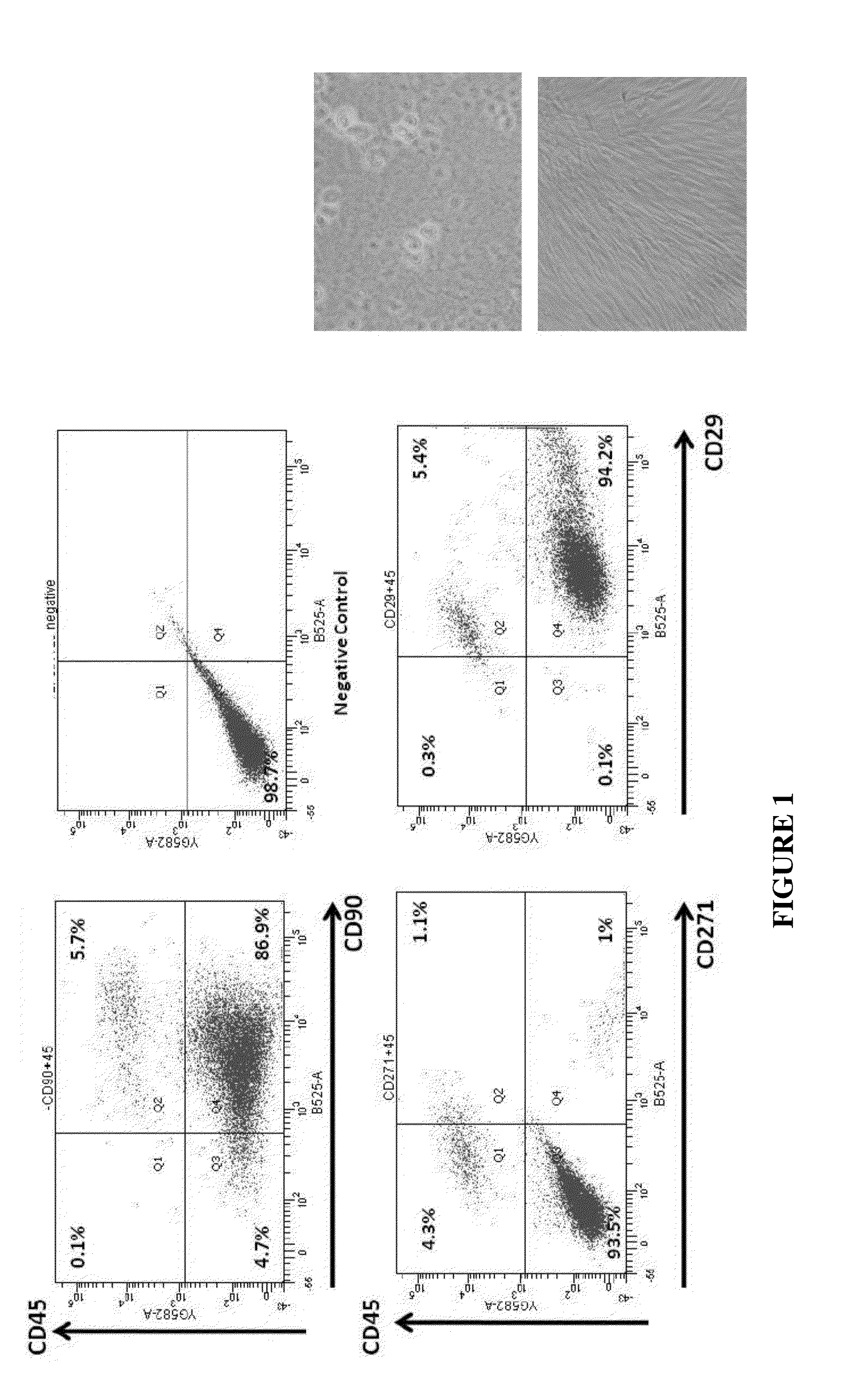
Stromal Vascular Fraction

[0125]The stromal vascular fraction (SVF) of adipose tissue is known to contain mesenchymal stem cells (MSC), T regulatory cells, endothelial precursor cells, preadipocytes, as well as anti-inflammatory M2 macrophages [104]. Accordingly, the SVF also contains blood cells from the capillaries supplying the fat cells. Safety of autologous adipose tissue implantation is supported by extensive use of this procedure in cosmetic surgery, as well as by ongoing studies using in vitro expanded adipose derived MSC (ADSC). Equine and canine studies demonstrating anti-inflammatory and regenerative effects of non-expanded SVF cells (non-cultured) have yielded promising results. Non-expanded SVF cells have been used successfully in accelerating healing of Crohn's fistulas and in treatment of multiple sclerosis [104]. Additionally, autologous SVF administration has been used commercially in over 3000 race horses for post-injury acceleration of healing, with published effic...

example 2

The Method for Extracting, Providing and Administrating the SVF

[0129]It is notable that the whole process of liposuction, SVF extraction and injection could take approximately 2 hours.

a—Liposuction

[0130]Conventional procedure of liposuction to extract abdominal fat will be performed at the operating room. In brief, after performing local anesthesia and administrating vasoconstrictor (while the fat tissue settles and becomes less swollen), an stab wound in abdomen will be made by a 11 blade scalpel and stainless steel cannulas or microcannulas will be inserted into subcutaneous tissue. Then 200 to 300 mL adipose tissue is pumped out and collected in a sterile 1 L storage bottles (Corning, N.Y., USA). The aspirate bottle will be kept in the ice packed box. For optimal results, only aspirate that has been obtained by tumescent liposuction method was used.

b—Adipose Tissue Preservation and Delivery:[0131]The SVF harvesting protocol which is used is based on the frequently applied methods...

example 3

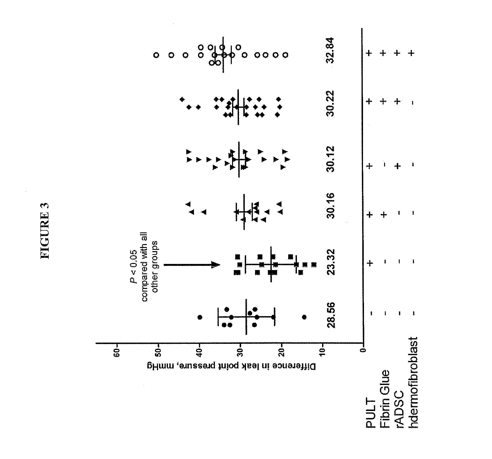
PUL Deficiency Causes SUI in Female Rats

[0191]A model of SUI in female rats has been developed and described [56,57]. In summary, these published data confirm the phenotype and functional similarities of PUL between humans and female rats to the level that PUL injury causes SUI in rats (FIG. 7 and FIG. 8). In addition, the feasibility of injection of the material (Fibrin Glue and MSC) in Canine Models of PUL deficiency has been tested (FIG. 9).


PUM

PropertyMeasurementUnit
Diameteraaaaaaaaaa
Stress optical coefficientaaaaaaaaaa
Biocompatibilityaaaaaaaaaa

Abstract

The invention relates to methods for the treatment of urinary incontinence (UI) and stress urinary incontinence (SUI) in both men and women by replacement of a damaged, absent or injured pubo-urethral ligament (PUL) via a simple injection of “Regenerative Glue.” For example, in the case of UI, the injection of “Regenerative Glue” at the PUL site will adhere the ventral surface of the urethra to the dorsal surface of the symphysis pubis (i.e. the space of Retzius), and further provide regenerative material for continuous replacement of deficient tissues and cells. “The Regenerative Glue” can be made from several commercially available materials including biocompatible glues, fibrin sealant or biocompatible gels; combined with Meschencymal stem cells (MSC) derived from bone marrow or adipose tissues.

Description


Claims


Application Information

Patent Timeline
Owner UNIVERSITY HOSPITALS OF CLEVELAND CLEVELAND
Why Eureka
Try Eureka