Sonogenic Stimulation of Cells

a cell and cell technology, applied in the field of cell and cell stimulation, can solve the problems of difficulty in delivering stimulus to the target neurons present in the deeper brain regions, and achieve the effects of reducing one or more symptoms of a disease, reducing viral replication, and reducing enzyme activity

Active Publication Date: 2016-08-04
SALK INST FOR BIOLOGICAL STUDIES
View PDF5 Cites 47 Cited by
  • Summary
  • Abstract
  • Description
  • Claims
  • Application Information

AI Technical Summary

Benefits of technology

[0039]An “effective amount” is an amount sufficient to accomplish a stated purpose (e.g. achieve the effect for which it is administered, treat a disease, reduce enzyme activity, reduce one or more symptoms of a disease or condition, reduce viral replication in a cell). An example of an “effective amount” is an amount sufficient to contribute to the treatment, prevention, or reduction of a symptom or symptoms of a disease, which could also be referred to as a “therapeutically effective amount.” A “reduction” of a symptom or symptoms (and grammatical equivalents of this phrase) means decreasing of the severity or frequency of the symptom(s), or elimination of the symptom(s). A “prophylactically effective amount” of a drug is an amount of a drug that, when administered to a subject, will have the intended prophylactic effect, e.g., preventing or delaying the onset (or reoccurrence) of an injury, disease, pathology or condition, or reducing the likelihood of the onset (or reoccurrence) of an injury, disease, pathology, or condition, or their symptoms. The full prophylactic effect does not necessarily occur by administration of one dose, and may occur only after administration of a series of doses. Thus, a prophylactically effective amount may be administered in one or more administrations. An “activity decreasing amount,” as used herein, refers to an amount of antagonist required to decrease the activity of an enzyme or protein (e.g. Tat, Rev) relative to the absence of the antagonist. A “function disrupting amount,” as used herein, refers to the amount of antagonist required to disrupt the function of an enzyme or protein relative to the absence of the antagonist. The exact amounts will depend on the purpose of the treatment, and will be ascertainable by one skilled in the art using known techniques (see, e.g., Lieberman, Pharmaceutical Dosage Forms (vols. 1-3, 1992); Lloyd, The Art, Science and Technology of Pharmaceutical Compounding (1999); Pickar, Dosage Calculations (1999); and Remington: The Science and Practice of Pharmacy, 20th Edition, 2003, Gennaro, Ed., Lippincott, Williams & Wilkins).

Problems solved by technology

While these methods have revealed insights into circuit computations in a number of model systems including mice, they suffer from one drawback: difficulty in delivering stimulus to the target neurons that are present in deeper brain regions.

Method used


Image

  • Sonogenic Stimulation of Cells
  • Sonogenic Stimulation of Cells
  • Sonogenic Stimulation of Cells

Examples


example 1

Imaging Setup Delivers Ultrasound Waves to Animals

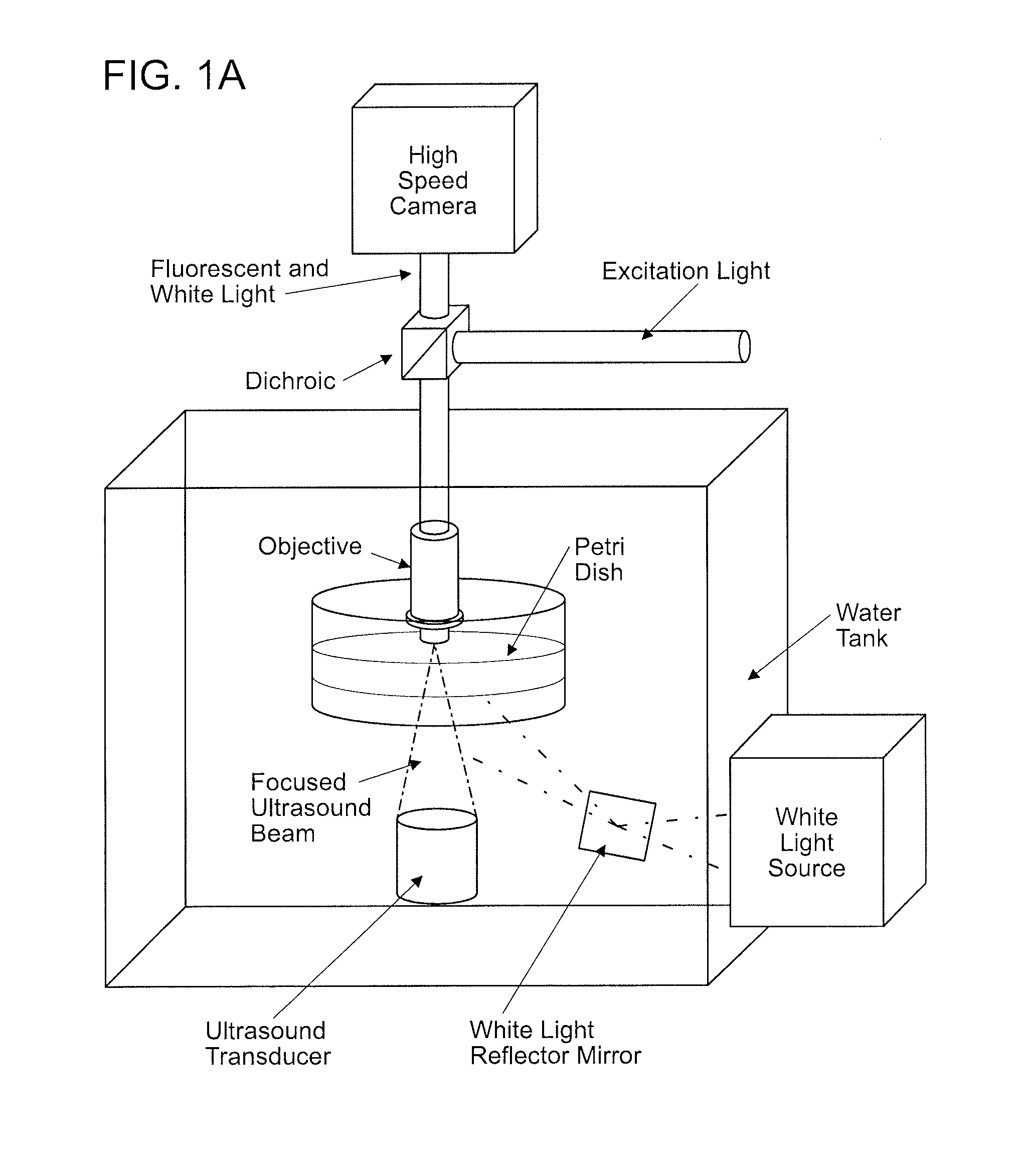
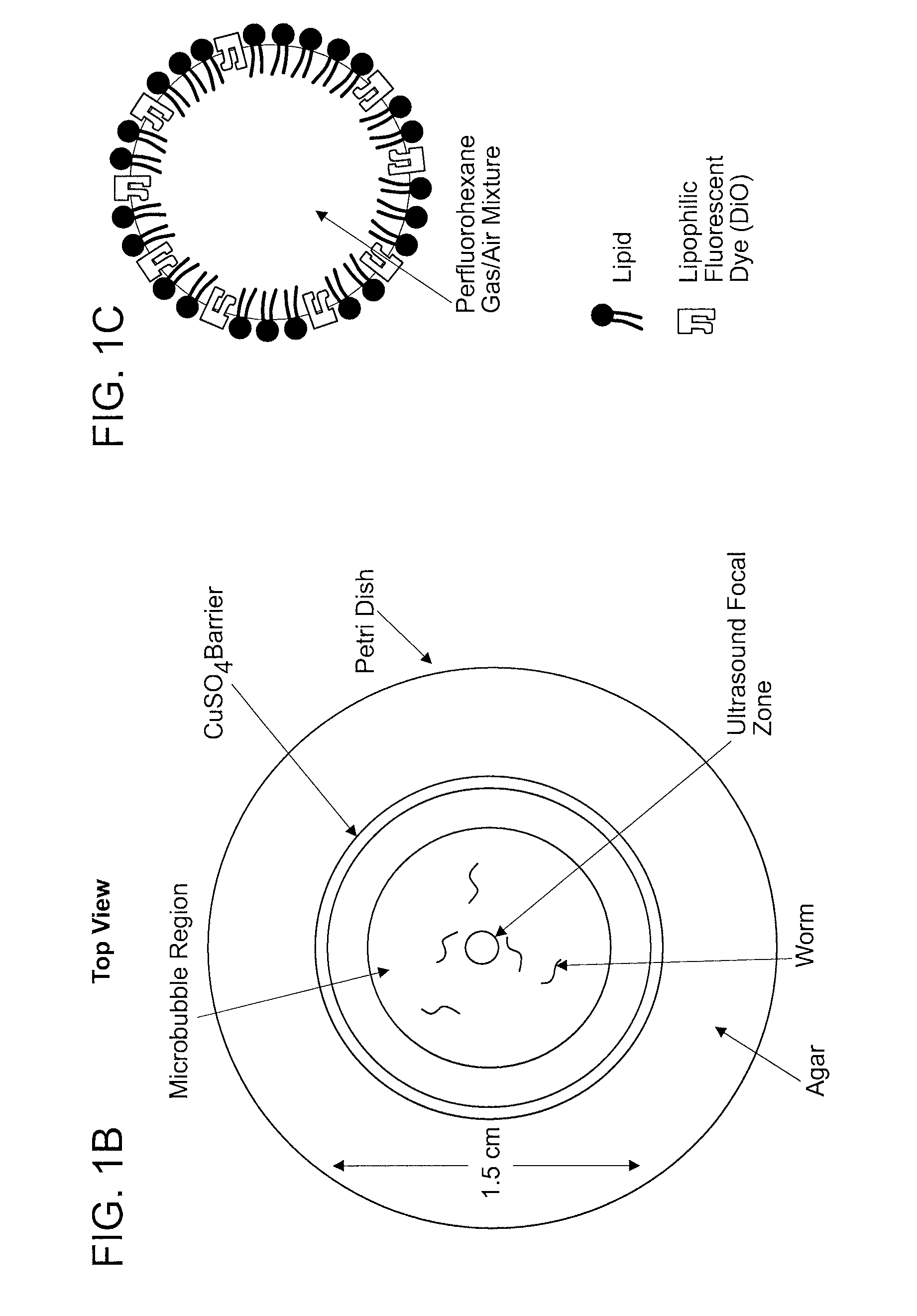
[0079]To investigate the role of ultrasound on C. elegans behavior, Applicants developed a novel imaging setup (FIG. 1A). Low intensity ultrasound was generated from a transducer and focused onto an agar plate where animals were corralled into a small area using a copper solution (FIG. 1B). Applicants' setup allowed for the ultrasound wave to be focused to a 1 mm diameter circular area at the agar surface (FIGS. 8A-8C). The whole setup was placed in a large tank filled with water to facilitate uniform transduction of the ultrasound wave. Previous studies have shown that at high ultrasound intensities (>2.5 MPa) water vapor bubbles would form spontaneously and collapse rapidly, initiating shockwaves that would compromise the integrity of cell membranes (termed “cavitation”) (C. K. Holland and R. E. Apfel, J Acoust Soc Am 88 (5), 2059 (1990); S. Bao, B. D. Thrall, and D. L. Miller, Ultrasound Med Biol 23 (6), 953 (1997)). Applicants co...

example 2

Microbubbles Amplify the Mechanical Deformation of the Ultrasound Wave

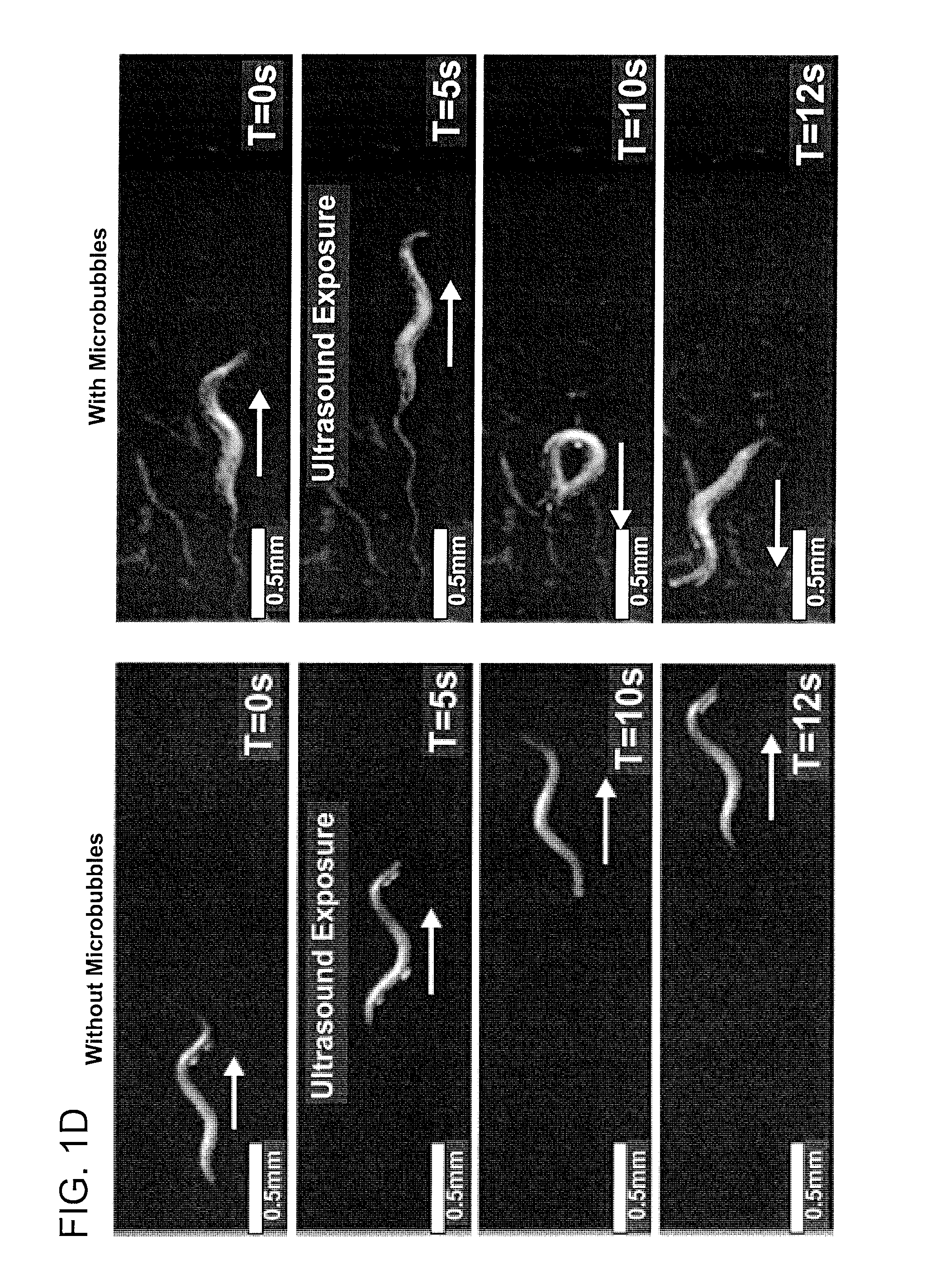
[0082]To amplify the ultrasound wave, Applicants included gas-filled microbubbles in Applicants' assay (FIG. 1C). Previous studies have shown that the majority of the ultrasound energy propagates through water and soft tissue as a longitudinal wave with alternating compression and rarefaction phases. These two phases create pressures that are alternately higher and lower than the ambient pressure level respectively. Applicants designed the microbubbles to respond to the mechanical deformations induced by an ultrasound pulse. Applicants filled the microbubbles with a stabilizing mixture of perfluorohexane and air that allows the compression and rarefaction phases of the ultrasound wave to shrink and expand the microbubbles from one half to four times their original diameters in a process known as stable cavitation. This occurs at the driving frequency of the underlying ultrasound pulse. Applicants found that animal...

example 3

TRP-4 Stretch Sensitive Ion Channels Sensitize Neurons to Ultrasound

[0083]Applicants hypothesized that ultrasound is a mechanical stimulus that require specific mechanotransduction channels to transduce the signals in individual neurons. Applicants tested the ability of TRP-4, a pore forming cation-selective mechanotransduction channel (L. Kang, J. Gao, W. R. Schafer et al., Neuron 67 (3), 381 (2010); W. Li, Z. Feng, P. W. Sternberg et al., Nature 440 (7084), 684 (2006)), to transduce this ultrasound induced mechanical stimulus. This channel is specifically expressed in a few C. elegans neurons, the four CEPs (CEPDL, CEPDR, CEPVL and CEPVR) and the two ADE (ADEL and ADER) dopaminergic neurons and the DVA and DVC interneurons (L. Kang, J. Gao, W. R. Schafer et al., Neuron 67 (3), 381 (2010); W. Li, Z. Feng, P. W. Sternberg et al., Nature 440 (7084), 684 (2006)). TRP-4 is both necessary and sufficient to generate mechanoreceptor currents in CEP neurons. Applicants found that animals m...


PUM

PropertyMeasurementUnit
frequencyaaaaaaaaaa
pressureaaaaaaaaaa
diameteraaaaaaaaaa

Abstract

The invention provides compositions featuring TRP-4 polypeptides and polynucleotides, methods for expressing such polypeptides and polynucleotides in a cell type of interest, and methods for inducing the activation of the TRP-4 polypeptide in neurons and other cell types using ultrasound.

Description

CROSS REFERENCE TO RELATED APPLICATIONS[0001]This application claims benefit of U.S. Provisional Application Ser. No. 62 / 054,600, filed Sep. 24, 2015, the contents of which are incorporated herein by reference.STATEMENT OF RIGHTS TO INVENTIONS MADE UNDER FEDERALLY SPONSORED RESEARCH[0002]This work was supported by the following grants from the National Institutes of Health, Grant Nos: NIH R01MH096881-03. The government has certain rights in the invention.BACKGROUND OF THE INVENTION[0003]Understanding how neural circuits generate specific behaviors requires an ability to identify the participating neurons, record and perturb their activity patterns. The best-understood motor circuit, the crab stomatogastric ganglion (STG) has benefited from electrophysiological access to well-defined cell types as well as an ability to manipulate them. A number of approaches have been developed for manipulating neuronal activity using light (optogenetics) or small molecules. While these methods have ...

Claims


Application Information

Patent Timeline
Patent Type & Authority Applications(United States)
IPC IPC(8): A61K41/00A61K38/17A61N7/00C12N13/00
CPCA61K41/0028A61N7/00A61K38/177C12N13/00A61K41/0033C07K14/705A61K38/00A61N2007/0026A61B8/00G01N29/00
Inventor CHALASANI, SREEKANTH H.IBSEN, STUART
Owner SALK INST FOR BIOLOGICAL STUDIES
Why Eureka
Try Eureka