Ultrasound image diagnostic apparatus

Active Publication Date: 2015-03-19
FUJIFILM CORP
View PDF10 Cites 2 Cited by
  • Summary
  • Abstract
  • Description
  • Claims
  • Application Information

AI Technical Summary

Benefits of technology

[0020]According to the ultrasound image diagnostic apparatus of the invention having the foregoing configuration, it is possible to shorten the time required for measu

Problems solved by technology

However, in the case where a region of interest is set in an ultrasound image, a tissue or lesion the operator desires to observe may be displaced from the set region of interest due to m

Method used


Image

  • Ultrasound image diagnostic apparatus
  • Ultrasound image diagnostic apparatus
  • Ultrasound image diagnostic apparatus

Examples


Example

[0025]An ultrasound image diagnostic apparatus of the invention is described in detail below with reference to a preferred embodiment shown in the accompanying drawings.

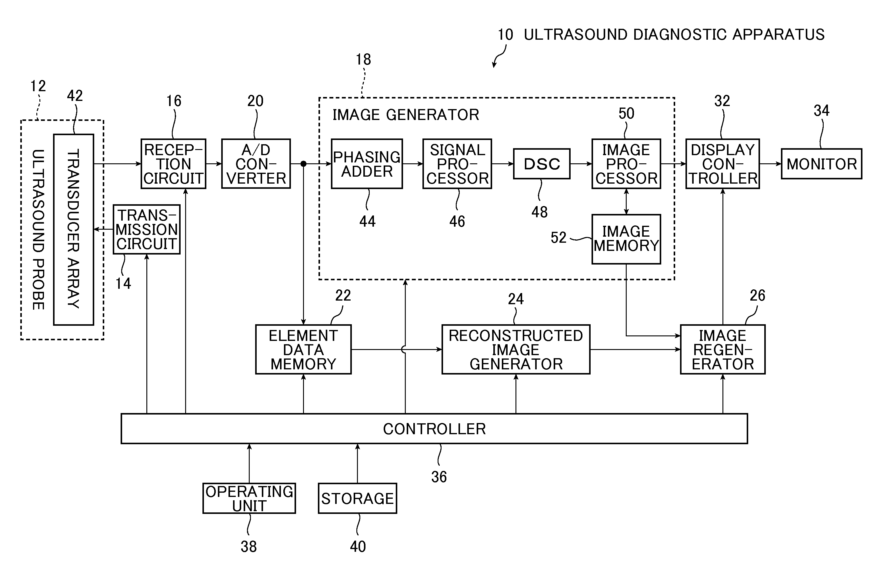
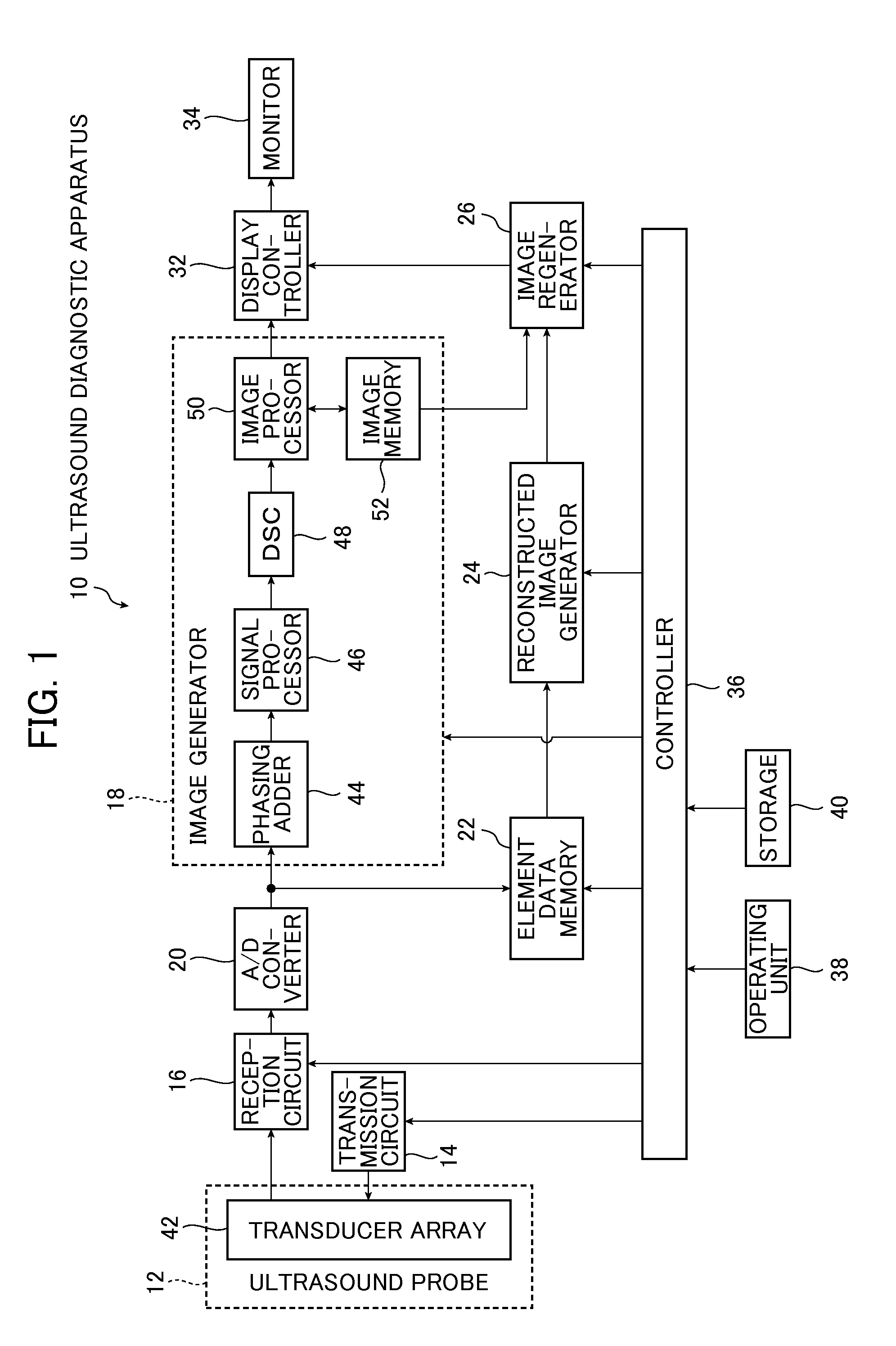
[0026]FIG. 1 is a block diagram conceptually showing an example of the configuration of the ultrasound image diagnostic apparatus (hereinafter also referred to as “ultrasound diagnostic apparatus”) of the invention.

[0027]An ultrasound diagnostic apparatus 10 includes an ultrasound probe 12, a transmission circuit 14 and reception circuit 16 connected to the ultrasound probe 12, an A / D converter 20, an image generator 18, an element data memory 22, a reconstructed image generator 24, an image regenerator 26, a display controller 32, a monitor 34, a controller 36, an operating unit 38 and a storage 40.

[0028]The ultrasound probe 12 includes a transducer array 42 of type used in general ultrasound diagnostic apparatuses.

[0029]The transducer array 42 comprises a plurality of ultrasound transducers arranged one-dimensional...


PUM


Abstract

The present invention aims at providing an ultrasound image diagnostic apparatus capable of shortening the time required for measurement and calculation of an optimal sound velocity and obtaining an ultrasound image having excellent image quality at a tissue or lesion the operator desires to observe. There are provided an element data memory storing element data; a region-of-interest setter setting a region of interest; an optimal sound velocity calculator calculating an optimal sound velocity at the region of interest using the element data corresponding to the region of interest; a reconstruction region setter setting a reconstruction region that contains the region of interest and is larger than the region of interest; and an image reconstructor generating a reconstructed image by reconstructing the ultrasound image in the reconstruction region based on the optimal sound velocity.

Description

CROSS REFERENCE TO RELATE APPLICATIONS[0001]This application is a continuation application of International Application No. PCT / JP2013 / 064038 filed on May 21, 2013, which claims priority under 35 U.S.C. 119(a) to patent application Ser. No. 2012-120011 filed in Japan on May 25, 2012, all of which are hereby expressly incorporated by reference into the present application.BACKGROUND OF THE INVENTION[0002]The present invention relates to an ultrasound image diagnostic apparatus that generates an ultrasound diagnostic image for use in diagnosis by imaging an organ and the like in a living body through transmission and reception of ultrasonic waves.[0003]Conventionally, ultrasound diagnostic apparatuses that use ultrasound images have been put to practical use in the medical field. In general, this type of ultrasound diagnostic apparatus comprises an ultrasound probe having a built-in transducer array and an apparatus body connected to the ultrasound probe. The ultrasound probe transmit...

Claims


Application Information

Patent Timeline
IPC IPC(8): A61B8/08A61B8/00A61B8/14
CPCA61B8/5215A61B8/14A61B8/5269A61B8/469A61B8/4488G16H50/20A61B8/463
Inventor KATSUYAMA, KIMITO
Owner FUJIFILM CORP
Why Eureka
Try Eureka