Method and System for 3D Reconstruction of X-ray CT Volume and Segmentation Mask from a Few X-ray Radiographs

a radiograph and ct volume technology, applied in tomography, image enhancement, instruments, etc., can solve the problems of more time-consuming and expensive acquiring a ct scan compared to a standard x-ray scan

Active Publication Date: 2019-07-18
SIEMENS HEALTHCARE GMBH
View PDF1 Cites 24 Cited by
  • Summary
  • Abstract
  • Description
  • Claims
  • Application Information

AI Technical Summary

Benefits of technology

[0004]The present invention provides a method and system for automated computer-based reconstruction of 3D computed tomography (CT) volumes from a small number of X-ray radiograph...

Problems solved by technology

Moreover, acquiring a CT scan is more time consuming...

Method used


Image

  • Method and System for 3D Reconstruction of X-ray CT Volume and Segmentation Mask from a Few X-ray Radiographs
  • Method and System for 3D Reconstruction of X-ray CT Volume and Segmentation Mask from a Few X-ray Radiographs
  • Method and System for 3D Reconstruction of X-ray CT Volume and Segmentation Mask from a Few X-ray Radiographs

Examples


Embodiment Construction

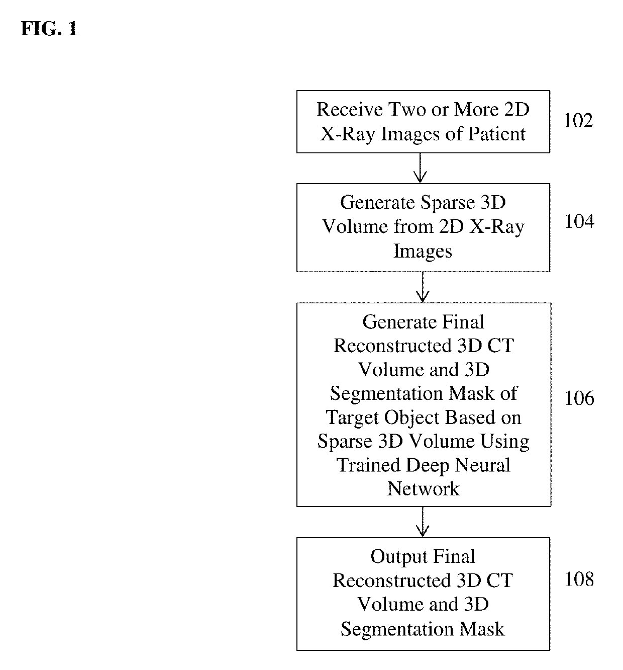
[0013]The present invention relates to a method and system for automated computer-based reconstruction of 3D computed tomography (CT) volumes and generation of 3D segmentation masks from a small number of X-ray radiographs. Embodiments of the present invention are described herein to give a visual understanding of the method for automated reconstruction of 3D CT volumes and generation of 3D segmentation masks. A digital image is often composed of digital representations of one or more objects (or shapes). The digital representation of an object is often described herein in terms of identifying and manipulating the objects. Such manipulations are virtual manipulations accomplished in the memory or other circuitry / hardware of a computer system. Accordingly, is to be understood that embodiments of the present invention may be performed within a computer system using data stored within the computer system.

[0014]Embodiments of the present invention provide automated computer-based recons...


PUM


Abstract

A method and apparatus for automated reconstruction of a 3D computed tomography (CT) volume from a small number of X-ray images is disclosed. A sparse 3D volume is generated from a small number of x-ray images using a tomographic reconstruction algorithm. A final reconstructed 3D CT volume is generated from the sparse 3D volume using a trained deep neural network. A 3D segmentation mask can also be generated from the sparse 3D volume using the trained deep neural network.

Description

BACKGROUND OF THE INVENTION[0001]The present invention relates to automated reconstruction of a 3D computed tomography (CT) volume from 2D X-ray radiographs, and more particularly, to automated reconstruction of a 3D CT volume and a 3D segmentation mask from a small number of X-ray radiographs.[0002]X-ray computed tomography (CT) is widely used for obtaining images of different parts of the body which are more detailed than standard X-rays. In computed tomography, many X-ray images are taken from various angles. All of these X-ray images are then processed using a tomographic reconstruction algorithm to generate the 3D CT volume data, which can then be view as images in the axial, coronal, sagittal planes.[0003]CT is regarded as a moderate to high radiation diagnostic technique. Moreover, acquiring a CT scan is more time consuming and expensive compared to acquiring a standard X-ray scan. Therefore, in order to decrease patients' exposure to radiation and reduce time and cost, it wo...

Claims


Application Information

Patent Timeline
IPC IPC(8): A61B6/03G06T7/11G06T11/00G06T7/00A61B6/00
CPCA61B6/032G06T7/11G06T11/005G06T7/0012A61B6/52G06T5/005G06T2207/10081G06T2200/04G06T2207/30004G06T17/00G06T2207/20084G06T2207/20081G06T11/003G06T2211/436
Inventor ZHOU, SHAOHUA KEVINNANDURI, SRI VENKATA ANIRUDHPARK, JIN-HYEONGLIAO, HAOFU
Owner SIEMENS HEALTHCARE GMBH
Why Eureka
Try Eureka