Apparatus for asymmetric dual-screen digital radiography

a dual-screen digital radiography and apparatus technology, applied in the field of digital radiography, can solve the problems of double exposure technique sensitive to patient motion artifacts, misregistration between the two images, degrade the subtraction image, etc., and achieve the effect of high detective quantum efficiency, sharper images, and higher x-ray absorption

Active Publication Date: 2008-01-17
CARESTREAM HEALTH INC
View PDF23 Cites 125 Cited by
  • Summary
  • Abstract
  • Description
  • Claims
  • Application Information

AI Technical Summary

Benefits of technology

[0053]The above-described asymmetric dual-screen digital radiography apparatus have various advantages over single-screen digital radiography apparatus. The present invention provides a higher detective quantum efficiency (DQE) which is a key metric for evaluating image quality. The higher MTF of the apparatus of the present invention yield sharper images. The higher X-ray absorption yields higher detector speed. The lower noise levels of the exemplary apparatus of the present invention give less quantum mottle. The higher detective quantum efficiency of the embodiments of the present invention provide higher overall image quality. Furthermore, the use of a pair of asymmetric screens in an indirect DR apparatus significantly eases the conflict in the design of an X-ray phosphor screen to maintain simultaneously both the X-ray absorption (which in general requires a screen with increased thickness) and the spatial resolution (which in general requires a screen with decreased thickness). Moreover, the use of a flexible substrate (e.g., metal foil, plastic sheet, or combinations thereof) for the flat panel imaging apparatus improves the mechanical strength and physical durability of the apparatus, and reduces the X-ray absorption loss due to the substrate.
[0054]The proposed single-exposure dual-energy digital radiography apparatus has various advantages over the prior art. There is no switching of the X-ray tube voltage required with such embodiments. There can be a reduction of patient motion misregistration artifacts in images. There can be lower X-ray tube loading, and lower X-ray exposure on the patient. In addition, higher detector performance in spatial resolution and signal-to-noise ratio can yield higher image quality and subsequent improvement in detection and characterization of diseases through dual-energy imaging. Moreover, there can be better integration of dual energy imaging into the current clinical workflow providing higher operation efficiency, without the needs of digitizing the film images (SF apparatus), scanning the imaging plates (CR apparatus), and double exposure on the patient (dual-exposure DR apparatus).
[0055]In at least one of the above-identified embodiments of the invention, instead of using two flat panels to capture the radiographic image, a single flat panel having a phosphor screen coated on the front side of the substrate which is thinner than the phosphor screen coated on the back side of the substrate is used in another exemplary embodiment of the present invention. A light blocking layer can be coated on each side of the substrate to minimize the crossover of light emitted in the phosphor screen on one side of the substrate to the photodiode on the other side of the substrate. The two screens are exposed to X-rays simultaneously and the photodiode on the front and back sides of the substrate detect the front and back image respectively. Subsequently, the two images are combined and processed to yield a higher quality image. Since the two images detected by the front and back screens are in mirror image relation to each other, one of the images should be inverted when the image processing is carried out.

Problems solved by technology

Generally, medical X-ray detectors employing a scintillating phosphor screen to absorb X-rays and produce light suffer the loss of spatial resolution due to lateral light diffusion in the phosphor screen.
Since a double exposure of the patient must be performed, and the switching of the X-ray tube voltage must take a finite time, the double exposure technique would be sensitive to patient motion artifacts and misregistration between the two images.
However, in clinical practice patient motion during the interval between the two radiographic exposures will degrade the subtraction image.
This would inherently reduce the throughout of the dual-energy radiographic procedure and reduce the image quality due to the image degradations caused by the film digitizer.
Therefore, in areas distant from the center of the X-ray beam, misregistration occurs.
Although the above-mentioned dual energy CR apparatus have provided new diagnostic information that is not furnished by the conventional screen-film apparatus, these apparatus have been hampered by poor subtraction effectiveness, workflow inconveniences, and Detective Quantum Efficiency (DQE) limitations of the CR technology.
Although these dual-exposure based DR apparatus have shown improvements in the detection and characterization of lung diseases, there still are issues such as X-ray tube loading, X-ray dosage on patient, and patient motion artifacts.

Method used


Image

  • Apparatus for asymmetric dual-screen digital radiography
  • Apparatus for asymmetric dual-screen digital radiography
  • Apparatus for asymmetric dual-screen digital radiography

Examples


Embodiment Construction

[0076]The following is a detailed description of the preferred embodiments of the invention, reference being made to the drawings in which the same reference numerals identify the same elements of structure in each of the several figures.

[0077]The present invention is directed to a digital radiography apparatus wherein an X-ray source projects an X-ray beam through an object to produce an X-ray image captured by a detecting member. In particular the present invention is directed to various embodiments related to indirect asymmetric dual-screen DR apparatus and single-exposure dual energy DR apparatus.

[0078]Indirect Asymmetric Dual-screen DR Apparatus

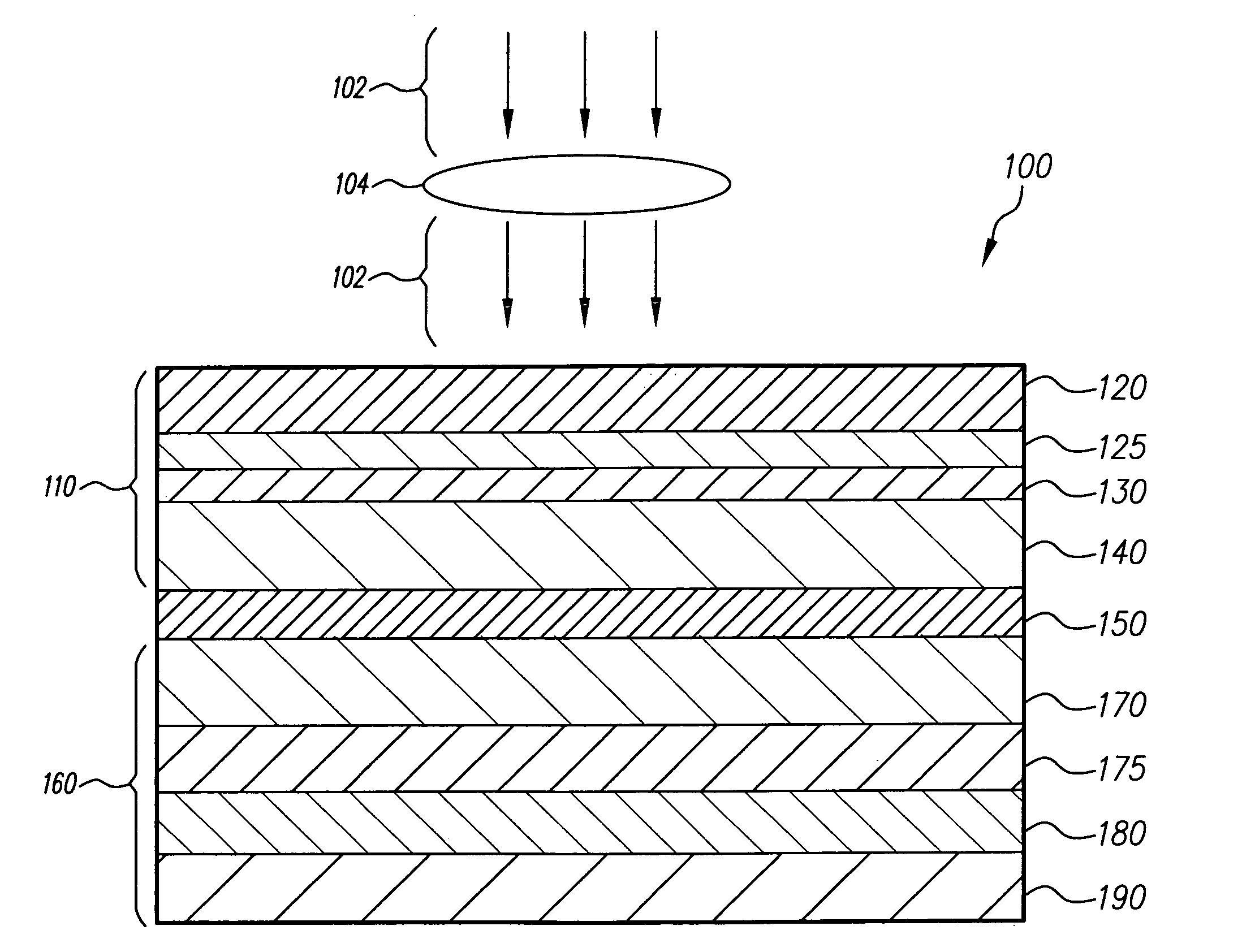
[0079]FIGS. 4-7 show diagrammatic views of a digital radiography apparatus in various embodiments in accordance with the present invention.

[0080]A first exemplary embodiment of the invention is illustrated in FIG. 4, where X-rays 102 are directed through object 104 to digital radiography imager 100 form an image. The digital radiography ...


PUM


Abstract

The present invention relates to radiographic imaging apparatus for taking X-ray images of an object. In various two-panel radiographic imaging apparatus configurations, a front panel and back panel have substrates, arrays of signal sensing elements and readout devices, and passivation layers. The front and back panels have scintillating phosphor layers responsive to X-rays passing through an object produce light which illuminates the signal sensing elements to provide signals representing X-ray images. The X-ray apparatus has means for combining the signals of the X-ray images to produce a composite X-ray image. Furthermore, the composition and thickness of the scintillating phosphor layers are selected, relative to each other, to improve the diagnostic efficacy of the composite X-ray image. Alternatively, a radiographic imaging apparatus has a single panel having arrays of signal sensing elements and readout devices and scintillating phosphor layers that are disposed on both sides of a single substrate. The present invention further relates to various embodiments of indirect dual-screen DR flat-panel imager apparatus that provide single-exposure dual energy imaging.

Description

FIELD OF THE INVENTION[0001]The invention relates generally to digital radiography, and in particular relates to the use of a plurality of asymmetric screens in digital radiographic flat-panel imagers to improve image quality.BACKGROUND OF THE INVENTION[0002]Generally, medical X-ray detectors employing a scintillating phosphor screen to absorb X-rays and produce light suffer the loss of spatial resolution due to lateral light diffusion in the phosphor screen. To reduce lateral light diffusion and maintain acceptable spatial resolution, the phosphor screens must be made sufficiently thin.[0003]The spatial resolution and X-ray detection ability of an imaging apparatus are often characterized by the modulation transfer function (MTF) and X-ray absorption efficiency, respectively. Thin phosphor screens produce better MTF at the expense of reduced X-ray absorption. Usually, the coating density and the thickness of the phosphor screen are used in the design tradeoff between spatial resolu...

Claims


Application Information

Patent Timeline
Patent Type & Authority Applications(United States)
IPC IPC(8): G01T1/24
CPCG01T1/2018G21K2004/04G21K4/00G01T1/20181G01T1/20184A61B6/00
Inventor YORKSTON, JOHNYIP, KWOK-LEUNGWOJCIK, TIMOTHY J.
Owner CARESTREAM HEALTH INC
Why Eureka
Try Eureka