A MRI brain tumor image segmentation method based on optimized U-net network model

A network model and image segmentation technology, applied in the field of brain tumor image segmentation, can solve the problems of medical diagnosis and treatment, increase the difficulty of signal-to-noise distinction, and reduce the accuracy of segmentation, and achieve fast segmentation, low cost, and improved accuracy. Effect

Inactive Publication Date: 2018-12-25
NORTHEASTERN UNIV
View PDF1 Cites 60 Cited by
  • Summary
  • Abstract
  • Description
  • Claims
  • Application Information

AI Technical Summary

Problems solved by technology

The segmentation results of some areas on the edge are very different from the gold standard, resulting in a decrease in segmentation accuracy, increasing the difficulty of signal-to-noise distinction, and having a serious impact on subsequent medical diagnosis and treatment.

Method used


Image

  • A MRI brain tumor image segmentation method based on optimized U-net network model
  • A MRI brain tumor image segmentation method based on optimized U-net network model
  • A MRI brain tumor image segmentation method based on optimized U-net network model

Examples


Embodiment 1

[0075] like Figure 5 Described, the present embodiment discloses a kind of MRI brain tumor image segmentation method based on optimized U-net network model, and described method comprises:

[0076] 101. Preprocessing the acquired multimodal MRI brain tumor image data.

[0077] The preprocessing process here is to remove the noise signal of the multimodal MRI brain tumor image data, and make it more closely match the algorithm of the model, so as to achieve the purpose of improving the quality of the final output image.

[0078] 102. Input the preprocessed multimodal MRI brain tumor image data into the trained U-net network model.

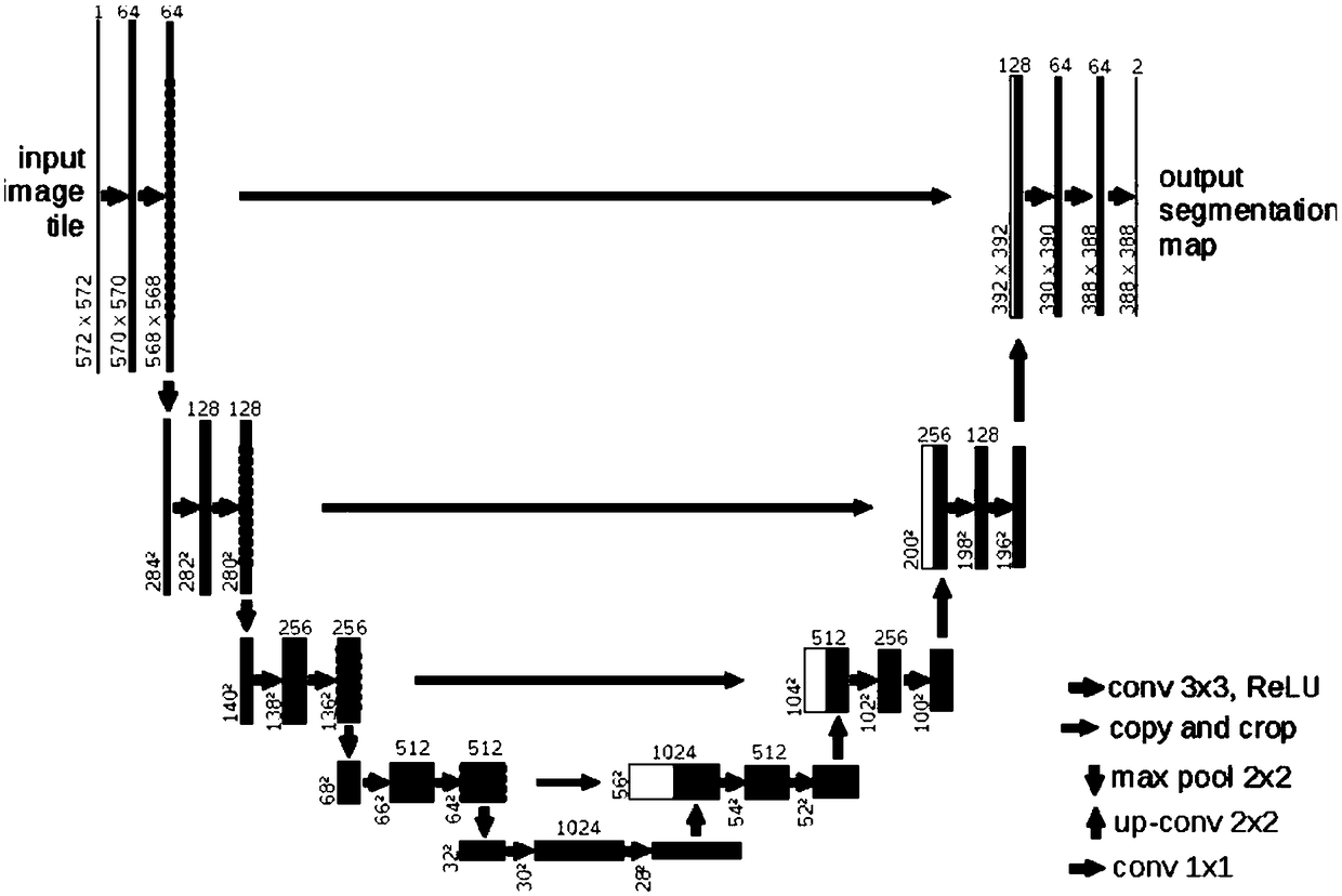
[0079] It should be noted that the U-net network model described here is an improved U-net network model, and it is a U-net network model after a large number of brain tumor images and cell structure images are trained.

[0080] 103. Obtain the multimodal MRI brain tumor image segmentation data output by the U-net network model.

[0081] Here, t...

Embodiment 2

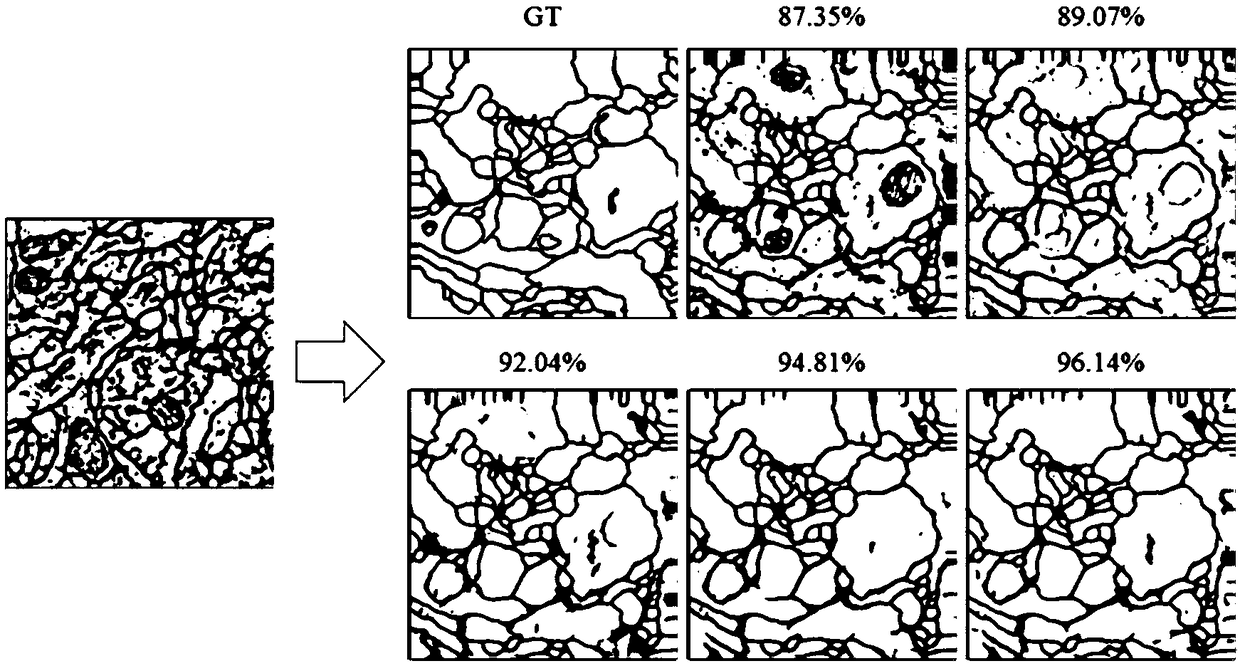
[0112] This embodiment combines the deep convolutional neural network and migration learning to improve the U-Net network model, and then uses the data set generated after simple data amplification, normalization and other preprocessing of the cell structure image to improve the U-Net network model. net network model for pre-training, and then use the multi-modal MRI brain tumor images (including HGG220 sets and LGG 54 sets) used in the BRATS2015 competition to fine-tune the network after preprocessing, and finally generate a multi-modal A deep learning model for segmentation of MRI brain tumor images. Framework flow chart such as Figure 8 shown.

[0113] Transfer learning: Deep learning has achieved major breakthroughs in computer vision tasks due to the use of very large datasets such as ImageNet to train networks. To effectively utilize smaller datasets, it is common to train a base model on a large dataset (such as ImageNet) and then fine-tune the learned model on a sec...


PUM


Abstract

The invention relates to a MRI brain tumor image segmentation method based on an optimized U-net network model. The method comprises the following steps: 101, preprocessing the acquired multimodal MRIbrain tumor image data; 102, inputting the preprocessed multimodal MRI brain tumor image data into the trained U-Net network model; 103, acquiring multi-modality MRI brain tumor image segmentation data output by a U-NET network model, wherein the multimodal MRI brain tumor image segmentation data output from the U-NET network model can preserve the image edge information to generate a complete segmented image feature map. The image segmentation method provided by the invention can not only preserve the image edge information and generate a complete characteristic map, but also improve the accuracy of image segmentation.

Description

technical field [0001] The invention belongs to the technical field of brain tumor image segmentation, in particular to an MRI brain tumor image segmentation method based on an optimized U-net network model. Background technique [0002] Local segmentation of brain lesions is crucial for medical diagnosis of brain tumors, surgical planning and prediction of disease progression. Deep neural networks have proven to be very promising algorithms for medical image segmentation. The deep learning algorithm does not need to manually specify the target features, the network can learn the data features from the gradually deepened convolutional layer, and can better extract features from the complex MRI image brain tumor data. The U-net network has achieved good results in the segmentation of cell structure images. However, after using the cell structure images to train the U-net network, after testing the test data, it is found that the segmentation results of some areas on the edge...

Claims


Application Information

Patent Timeline
Patent Type & Authority Applications(China)
IPC IPC(8): G06T7/12G06N3/04
CPCG06T7/12G06T2207/20081G06T2207/20084G06T2207/10088G06T2207/30096G06T2207/20192G06T2207/30016G06N3/045
Inventor 孝大宇张淑蕾王超高殿宇康雁
Owner NORTHEASTERN UNIV
Why Eureka
Try Eureka