Intravascular Diagnostic or Therapeutic Apparatus Using High-Intensity Pulsed Light

a high-intensity, pulsed light technology, applied in the field of intravascular diagnostic or therapeutic equipment, can solve the problems of inability to secure a sufficient time difficulty in allowing light to reach the region of the blood vessel targeted for diagnostics or therapy, and limited hemostasis time for avoiding ischemia

Inactive Publication Date: 2008-09-11
KEIO UNIV
View PDF10 Cites 61 Cited by
  • Summary
  • Abstract
  • Description
  • Claims
  • Application Information

AI Technical Summary

Benefits of technology

[0011]It is an object of the present invention to provide an intravascular diagnostic or therapeutic apparatus capable of removing blood in an intravascular lumen to be observed using a minimally invasive method. More specifically, the present invention is intended to provide a minimally invasive intravascular diagnostic or therapeutic apparatus which

Problems solved by technology

For this reason, it has been difficult to allow light to reach the region of the blood vessel targeted for diagnostics or therapy.
However, when the blood flow is stopped using the shut-off balloon, a hemostasis time for avoiding ischemia is limited and it is not possible to secure a sufficient time for diagnostics or therapy.
Moreover, when the field of view is secured by removing the blood in saline using a conventional angioscope, even a small amount of blood is mixed in, illumination light is diffused and reflected, which obstructs the field of view considerably.
This causes a vicious cycle of requiring further injection of a large amount of saline.
Furthermore, these conventional methods stop a blood flow or inject a large amount of foreign matters into the blood vessel, blocking the circulation of blood, which is an oxygen carrier, preventing a sufficient amount of oxygen from being supplied to peripherals and thus presenting a high level of invasiveness against an

Method used


Image

  • Intravascular Diagnostic or Therapeutic Apparatus Using High-Intensity Pulsed Light
  • Intravascular Diagnostic or Therapeutic Apparatus Using High-Intensity Pulsed Light
  • Intravascular Diagnostic or Therapeutic Apparatus Using High-Intensity Pulsed Light

Examples


example 1

[0121]The endoscope used in this example is shown in FIG. 5. As shown in FIG. 5, a small-diameter endoscope 22 was installed in a stainless steel sheath 23 having a length of approximately 3 cm and an inner diameter of 0.8 cm.

[0122]An image guide 20 and a lightguide 21 were placed in the small-diameter endoscope 22. A laser transmission fiber 19 is disposed along those guides and these were placed in a catheter sheath 18. In this case, the small-diameter endoscope 22, that is, the far ends of the image guide 20 and lightguide 21 were made to slightly protrude from the laser transmission fiber 19. Identical quartz optical fibers were used as the optical fibers for image pickup in the laser transmission optical fiber 19 and image guide 20. A plastic lightguide was used for the lightguide 21. The diameter of the laser transmission fiber 19 was approximately 0.6 mm and the diameter of the small-diameter endoscope 22 integrating the lightguide 21 and image guide 20 was approximately 0.7 ...

example 2

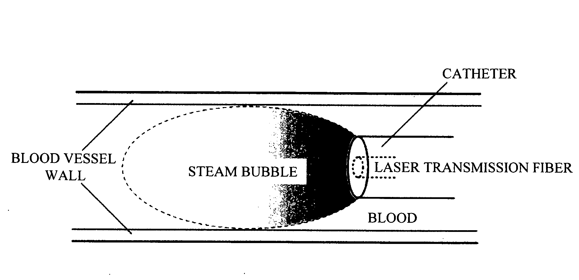
[0125]A silicon tube was filled with milk and the inner wall of the tube was observed using the endoscope of the present invention. The endoscope used was the same as that of Example 1. A silicon tube having an inner diameter of 3 mm was cut open, a piece of paper colored with water-resistant red ink was pasted inside and the silicon tube was closed again. Next, a far end part of a catheter sheath 18 in which a laser transmission optical fiber 19 of the endoscope, lightguide 21 and image guide 20 were disposed was inserted into the silicon tube and the tube was put in the milk so that the tube was filled with the milk. Next, pulsed laser was irradiated to generate water-vapor bubbles and images thereof were taken. The laser intensity at this time was 200 mJ / pulse or 450 mJ / pulse at the end of the laser irradiation fiber. The pulse width was approximately 200 μs. The images of the intravascular lumen delayed by a delay generator and taken by a CCD camera were displayed on a monitor a...

example 3

[0129]An aorta lumen of Japanese white rabbit was observed using the endoscope of the present invention. The structure of the endoscope used is in conformance with the endoscope shown in FIG. 5 used in Example 1, but a flash lamp excitation Ho:YAG laser (manufactured by Cyber Laser, model FLHY-1) was used for the laser generator. Furthermore, a fiber having a core diameter of 0.6 mm, outside diameter of 1.45 mm was used as the laser irradiation fiber and this fiber was used tied with an endoscope having an outside diameter of 1.3 mm (manufactured by au Medical Laboratory).

[0130]A 10 Fr. sheath was held to Japanese white rabbit aorta and the above described fiber tied with the endoscope was inserted therein.

[0131]The laser irradiation condition was 10 Hz, 400 mJ / pulse. As the control, images of the intravascular lumen were taken without laser irradiation.

[0132]FIG. 14 shows a photo of the intravascular lumen taken without laser irradiation and FIG. 15 shows a photo of the intravascul...


PUM


Abstract

An intravascular diagnostic or therapeutic apparatus capable of removing blood in an intravascular lumen to be observed using a minimally invasive method is provided. The apparatus includes high-intensity pulsed light generating means and high-intensity pulsed light transmitting means for transmitting high-intensity pulsed light, capable of irradiating the interior of a blood vessel with high-intensity pulsed light, producing water-vapor bubbles and temporarily removing the blood in the blood vessel.

Description

TECHNICAL FIELD[0001]The present invention relates to an intravascular diagnostic or therapeutic apparatus provided with high-intensity pulsed light irradiating means such as pulsed laser, which irradiates blood with the high-intensity pulsed light and generates water-vapor bubbles to enable diagnostics and therapy of an intravascular lumen. An example of such an apparatus is an angioscope which allows an observation of an intravascular lumen.BACKGROUND ART[0002]Conventionally, intravascular diagnostic or therapy has been conducted by inserting a catheter into a blood vessel using light for diagnostics or therapy. For example, a vessel lumen has been observed with an angioscope by irradiating illumination light. Furthermore, angioplasty or the like has been conducted by irradiating a vessel lumen with high-intensity pulsed light such as laser light. However, when light is irradiated into the blood vessel while blood exists in the blood vessel, light having a wavelength of visible li...

Claims


Application Information

Patent Timeline
IPC IPC(8): A61B18/20A61B1/00A61B1/015A61B1/04A61B1/12A61B18/24
CPCA61B1/015A61B18/24A61B1/3137A61B1/042A61B5/0084A61B5/02007A61B5/7285
Inventor ARAI, TSUNENORISUGA, ERIKOYAMASHITA, ERIKAFUTAMI, HIKARU
Owner KEIO UNIV
Why Eureka
Try Eureka