

Device for minimally invasive intravascular aortic valve extraction
a technology for aortic valves and extraction devices, which is applied in the field of devices for aortic valve extraction, can solve the problems of inability to fully open valves, and inability to perform extraction in medically complicated conditions, so as to prevent any risk of embolism and any risk
- Summary
- Abstract
- Description
- Claims
- Application Information
AI Technical Summary
Benefits of technology
Problems solved by technology
Method used
Image

Examples
Embodiment Construction
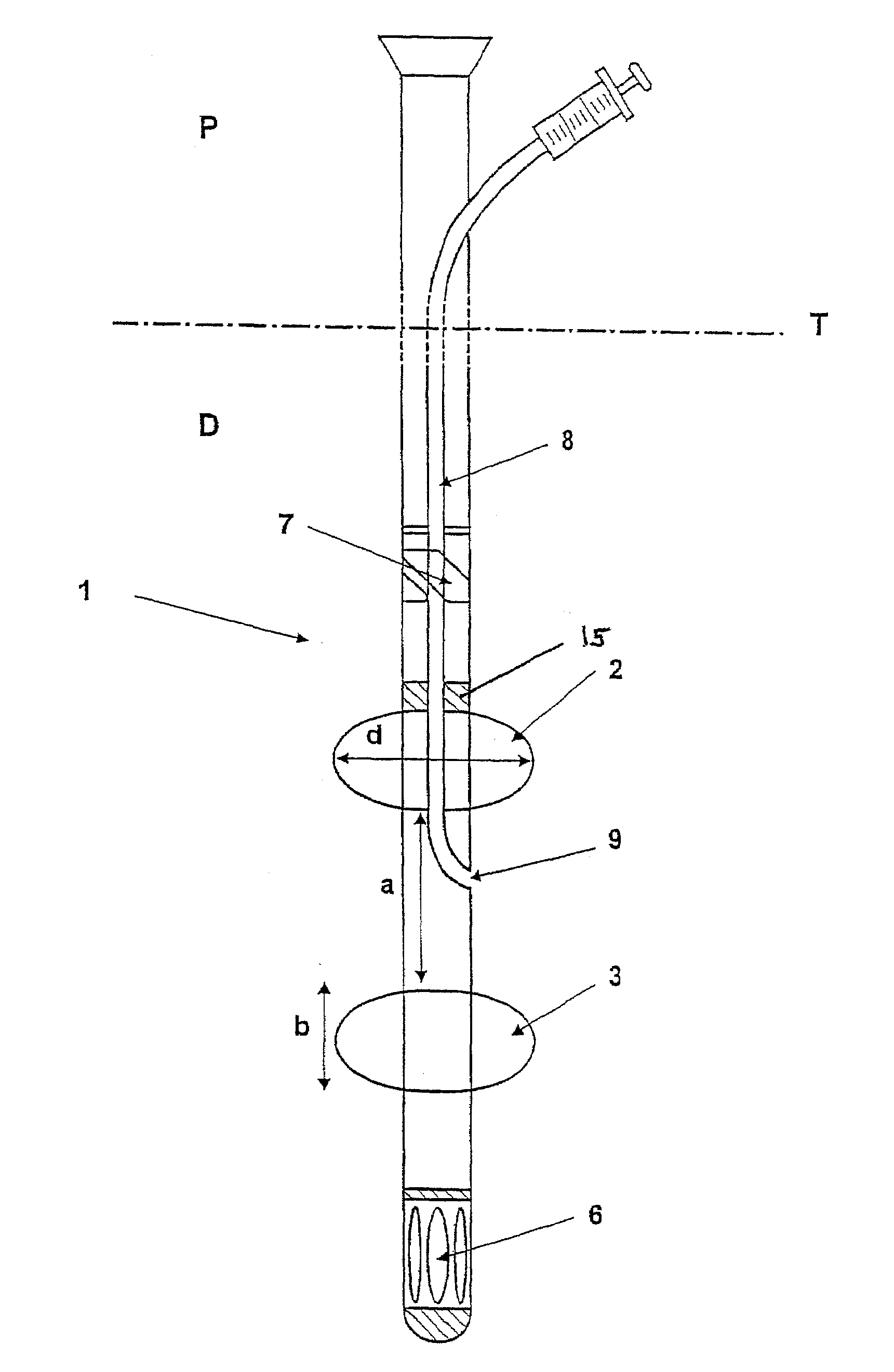
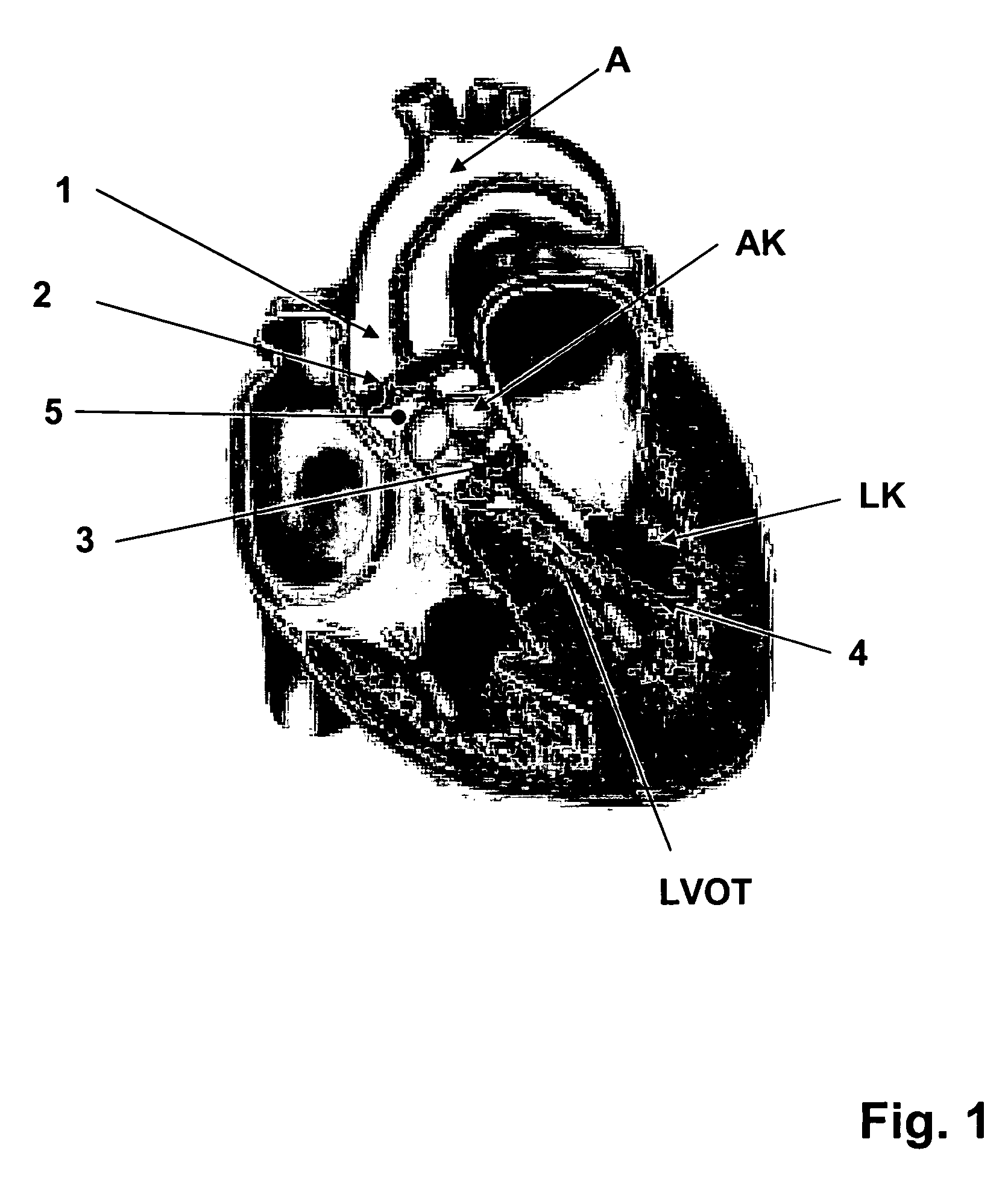
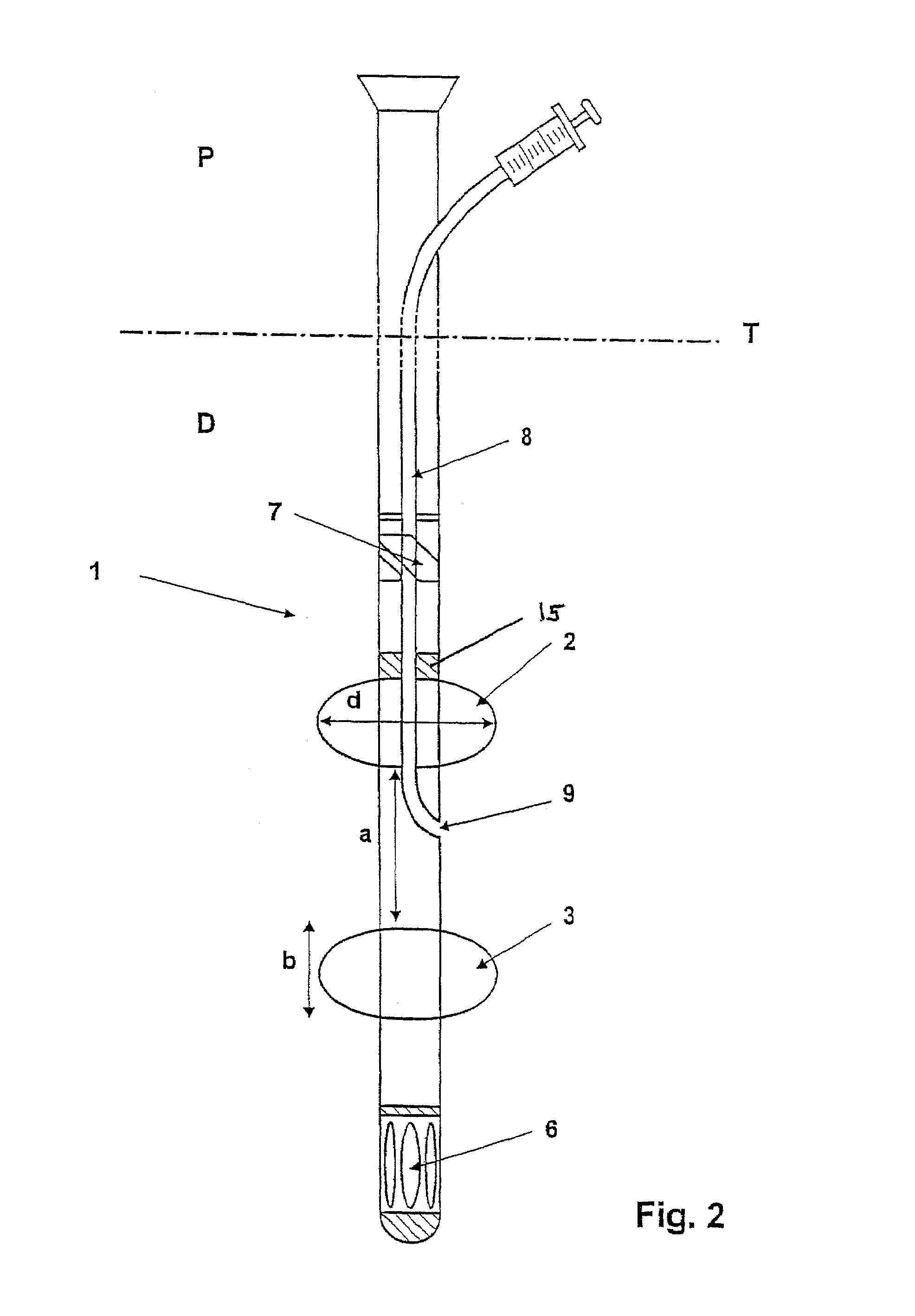
[0013]The object of the present invention, therefore, is to improve and provide a device for minimally invasive, intravascular aortic valve extraction which completely rules out any risk of embolism from tissue particles and / or calcium particles that might enter the blood stream during aortic valve extraction. Furthermore, the surgeon should be able to ablate diseased cardiac valve areas individually locally and selectively, preferably under direct optical observation of the diseased cardiac valve areas. In particular, handling of the device should be facilitated for the surgeon in such a manner that signs of fatigue which may set in due to the great concentration required to conduct the operation can be completely ruled out.
[0014]A further object of the present invention is to provide a respective method for minimally invasive, intravascular aortic valve extraction.
[0015]An element of the present invention is that a device for minimally invasive, intravascular aortic valve extracti...
PUM
 Abstract
Description
Claims
Application Information