Scanning/pre-scanning quality control of slides

a technology of quality control and scanning/prescanning, applied in the field of scanning/prescanning quality control of slides, can solve the problems of additional work, diagnostic pitfalls, and complete tissue uselessness

Pending Publication Date: 2022-08-18
VISIOPHARM AS
View PDF0 Cites 0 Cited by
  • Summary
  • Abstract
  • Description
  • Claims
  • Application Information

AI Technical Summary

Problems solved by technology

However, sometimes the presence of certain artefacts in a microscopic section can result in misinterpretations leading to diagnostic pitfalls.
Artefacts can even lead to complete uselessness of the tissue.
Since the pathologist may have to examine a large number of histology slides, any number of slides containing artefacts rendering the sample useless, results in additional work that could have been avoided, provided the artefacts had been detected at an earlier step in the process.
Performing a detailed image analysis of a plurality of histology slides is typically a very time-consuming process, and hence it is of interest to eliminate or at least minimize the number of useless slides, e.g. the ones containing artefacts, arriving at a pathologist for examination or analysis.

Method used


Image

  • Scanning/pre-scanning quality control of slides
  • Scanning/pre-scanning quality control of slides
  • Scanning/pre-scanning quality control of slides

Examples


example

[0064]An example of a digital scanner could be any of the Nanozoomer scanners from the company Hamamatsu, e.g. the Nanozoomer SQ, the Nanozoomer S60, the Nanozoomer S210, or the Nanozoomer S360.

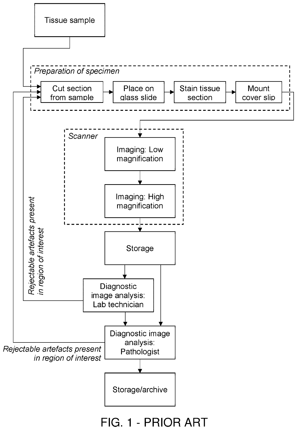
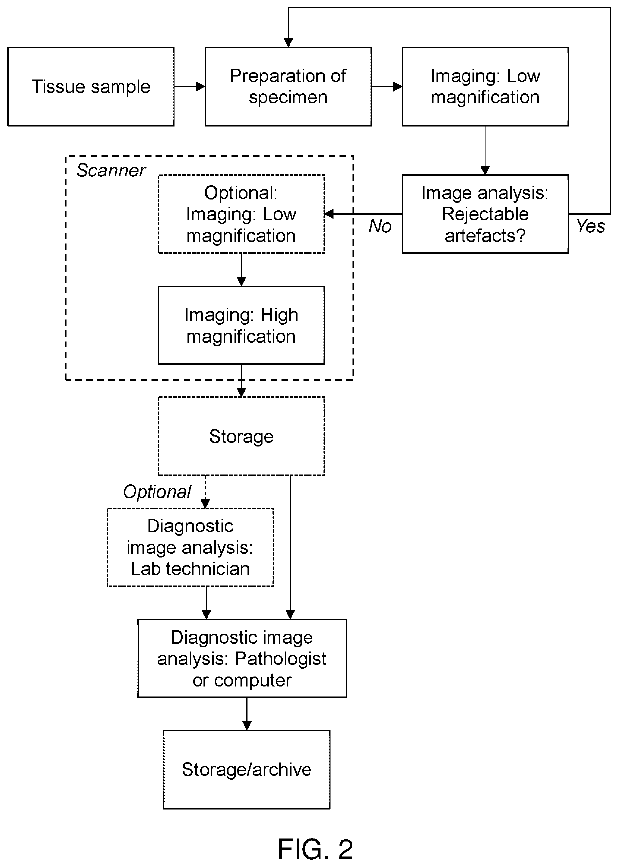
[0065]An example of a method for analysing a plurality of histology slides according to the present disclosure is shown in FIG. 2. In this example, the slides are analysed outside the scanner in order to evaluate whether each slide should be accepted or rejected. The imaging may be done by any camera capable of acquiring a low magnification image.

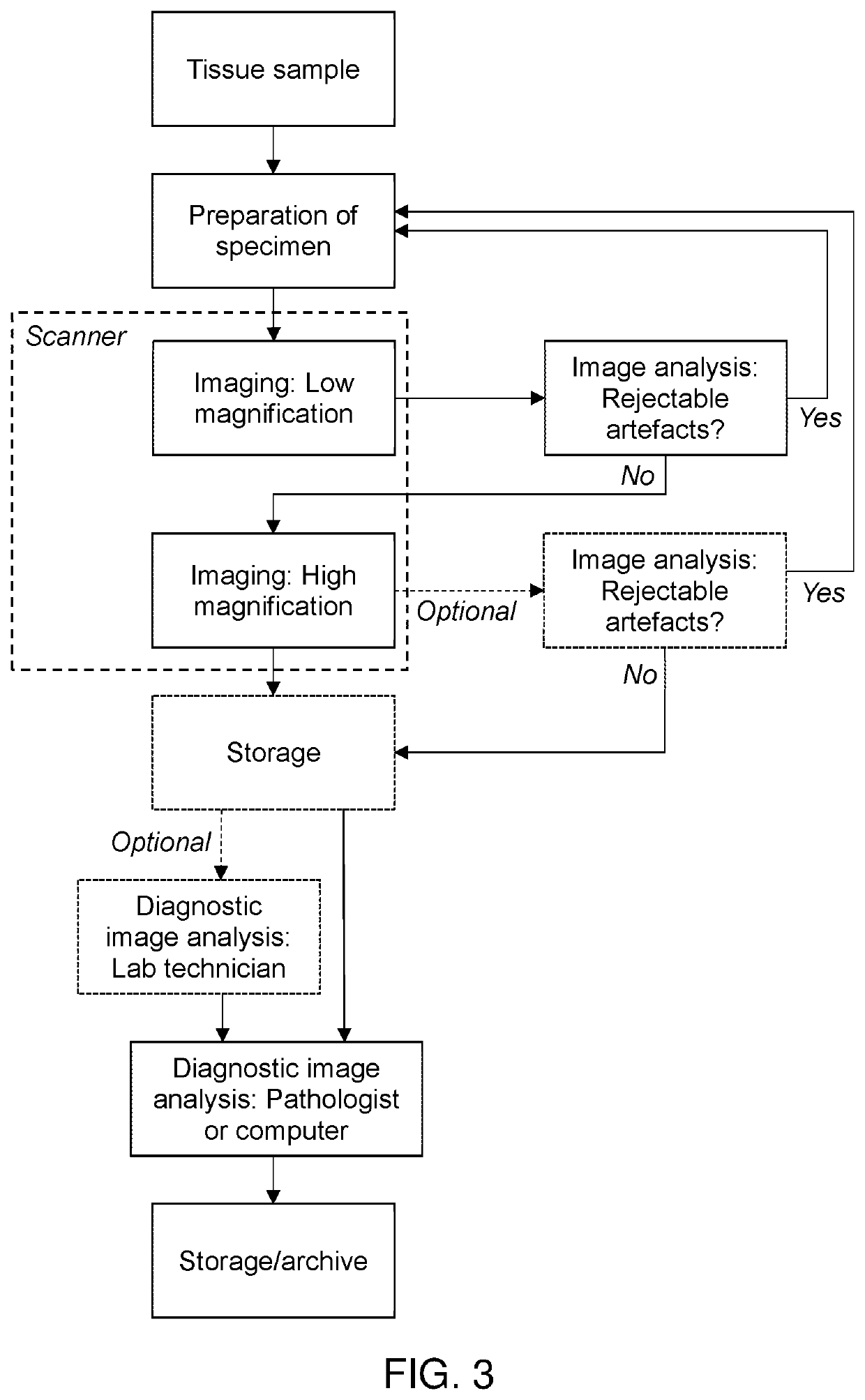
[0066]An example of a method for analysing a plurality of histology slides according to the present disclosure is shown in FIG. 3. In this example, the evaluation relating to rejectable artefacts is based on an image obtained inside the scanner, i.e. using the camera / lens provided by the scanner.


PUM


Abstract

A method of analyzing a plurality of histology slides is provided, wherein possible artefacts are detected at an early stage. This is achieved by including a preliminary imaging and image analysis step in the process flow; the step being executed preferably before any diagnostic assessment e.g. image analysis or manual reading is performed by a pathologist or a lab technician. Accordingly, the histology slides reaching the expert for image analysis are of a higher quality, since the slides are ideally free of artefacts. The method thus saves valuable time for the pathologist, since only artefact-free slides will be subject to a detailed analysis. Furthermore, the method minimizes the risk of misinterpretations leading to potentially false diagnoses. A deep learning model, is also disclosed, which is capable of automatically determining whether histopathological images are suitable for diagnostic and/or research assessment. A training of the deep learning model is also disclosed.

Description

CROSS-REFERENCE TO RELATED APPLICATIONS[0001]This application is the U.S. National Stage of PCT / EP2020 / 068037 filed Jun. 26, 2020, which claims priority to European Patent Application No. 19183338.3, filed Jun. 28, 2019, the content of both are incorporated herein by reference in their entirety.FIELD OF THE INVENTION[0002]The present disclosure relates to a method for analysing a plurality of histology slides as well as a method for preparing histology slides for analysis, and, based on an image of at least part of the tissue specimen, determining whether the slides comprise one or more rejectable artefacts. The present disclosure further relates to a method for training a deep learning model to recognize said artefacts.BACKGROUND OF INVENTION[0003]Histopathology refers to the microscopic examination of tissue in order to study the manifestations of disease. In clinical medicine, histopathology refers to the examination of a biopsy or surgical specimen by a pathologist, after the sp...

Claims


Application Information

Patent Timeline
Patent Type & Authority Applications(United States)
IPC IPC(8): G02B21/36G06T7/00G06V20/69G02B21/34G06V10/764
CPCG02B21/367G06T7/0012G06V20/695G06T2207/10056G06T2207/30024G06T2207/20081G06T2207/20084G02B21/34G01N1/30G06T2207/30168G06F18/24133G06V20/69G06V10/993G06V10/82G06V10/764
Inventor GRUNKIN, MICHAELHANSEN, JOHAN DORÉTHAGAARD, JEPPE
Owner VISIOPHARM AS
Why Eureka
Try Eureka