Method of Forming a Lesion in Heart Tissue

a heart tissue and tissue technology, applied in the field of thoracoscopic surgery of the cardiovascular system, can solve the problems of abnormal enlargement of the right chamber of the heart, abnormal enlargement of the body tissue supplied by the blood supply, and abnormal shunting of blood

Inactive Publication Date: 2006-09-05
HEARTPORT
View PDF108 Cites 364 Cited by
  • Summary
  • Abstract
  • Description
  • Claims
  • Application Information

AI Technical Summary

Benefits of technology

[0011]In a first aspect of the invention, a tubular access device is provided for accessing an interior chamber of a beating heart. The access device includes an elongated tubular body configured to extend percutaneously through an intercostal space between the ribs of the chest and through a muscular wall of the heart, and an inner lumen extending through the tubular body which provides an access channel into the heart. In an exemplary embodiment, the tubular access device has a length of at least 10 cm, and the inner lumen has a diameter of at least 5 mm. Preferably, the tubular access device is rigid to facilitate responsive and precise positionability from its proximal end.
[0040]By providing access to the interior of the heart without requiring a gross thoracotomy and without the need to induce cardioplegic arrest, the invention enables a variety of intracardiac procedures to be performed on a beating heart. In addition to septal defect repair and the electrophysiological procedures described above, these procedures may include repair of other types of congenital defects, transmyocardial laser revascularization, mitral, aortic, pulmonary, or tricuspid valve inspection and repair, pulmonary thrombectomy, intracardiac inspection, removal of growths, myxomas, neoplasms, hypertrophic obstructive cardiopmyopathy and vegetations, and other diagnostic and treatment procedures.

Problems solved by technology

These conditions may cause blood to abnormally shunt from the right side of the heart to the left side of the heart without being properly oxygenated in the lungs, so that the body tissues supplied by the blood are deprived of oxygen.
In addition, blood in the left side of the heart may shunt back to the right side through the defect rather than being pumped into the arterial system, causing abnormal enlargement of the right chambers of the heart.
The necessity of stopping the heart significantly heightens the risks attendant such procedures, particularly the risks of causing ischemic damage to the heart muscle, and of causing stroke or other injury due to circulatory emboli produced by aortic clamping and vascular cannulation.
In addition, the creation of a gross thoracotomy produces significant morbidity and mortality, lengthens hospital stay and subsequent recovery, increases costs, and worsens the pain and trauma suffered by the patient.
While intravascular approaches to the repair of congenital defects may provide certain advantages, the most significant of which is the elimination of the need for gross thoracotomy and cardioplegic arrest, these techniques have suffered from a number of problems.
One such problem is the difficulty in manipulating the artificial patches into position across a defect using only the proximal end of a long and flexible delivery catheter positioned through a tortuous right lumen.
Also problematic is the inadequacy of fixation of endovascularly-placed patches, creating a tendency of such patches to migrate or embolize after placement, which can allow blood to again shunt through the defect.
In addition, once such a patch has been placed and the delivery catheter detached from the patch, relocating and repositioning the patch with the catheter is difficult, if not impossible, and may require open surgical correction.
Moreover, in young children, the size of the peripheral vessels is extremely small, and damage to such vessels could have serious effects upon the growth of the child.
Thus, the size of the devices which can be introduced through such vessels is greatly limited.
However, endovascular mapping and ablation devices suffer from many of the same problems suffered by endovascular septal defect repair devices, including a lack of control and precise positionability from the proximal end of these highly flexible and elongated devices, the significant size constraints of peripheral vessels, and the inability to position the devices in all potentially diseased sites within the heart.

Method used


Image

  • Method of Forming a Lesion in Heart Tissue
  • Method of Forming a Lesion in Heart Tissue
  • Method of Forming a Lesion in Heart Tissue

Examples


Embodiment Construction

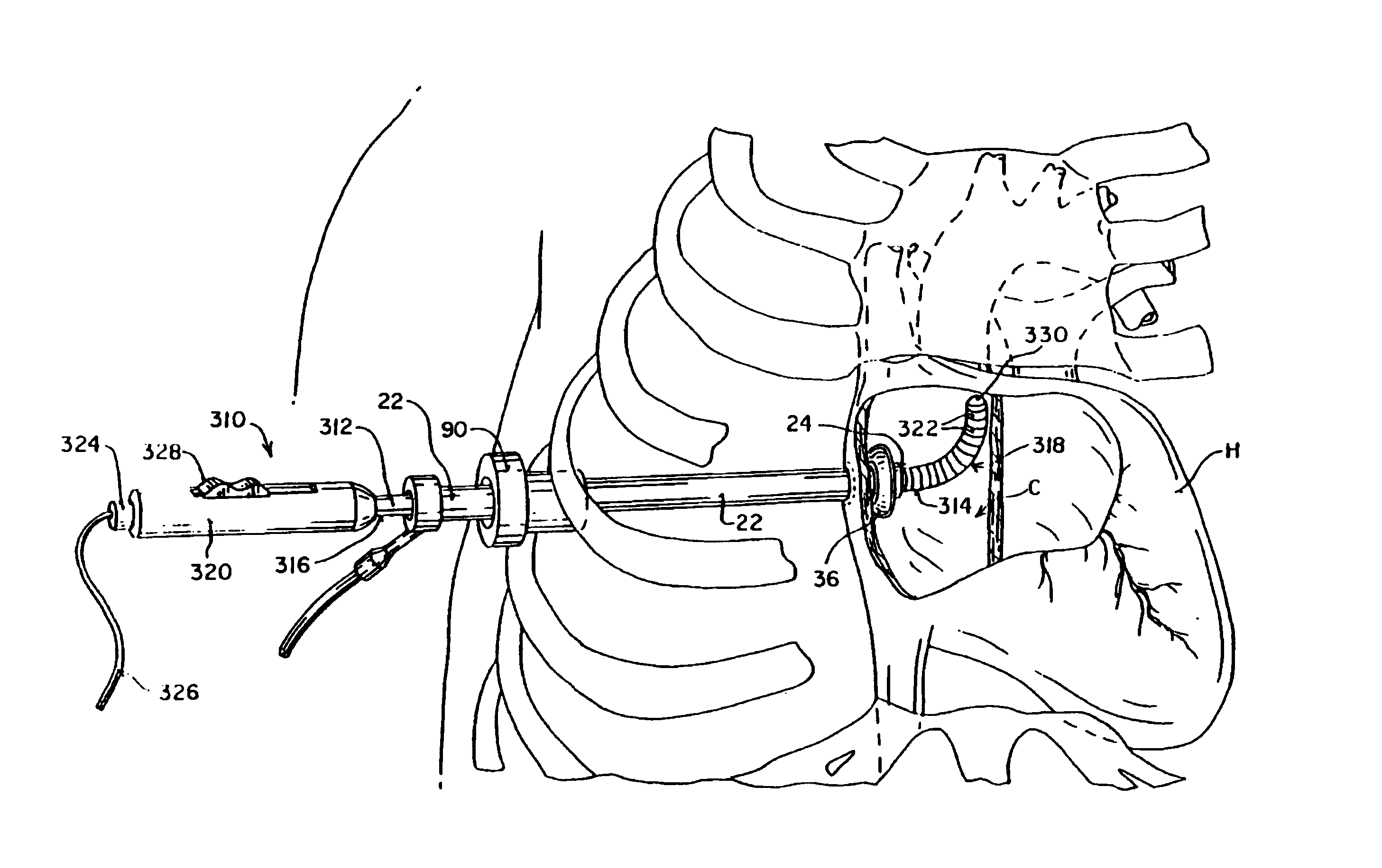
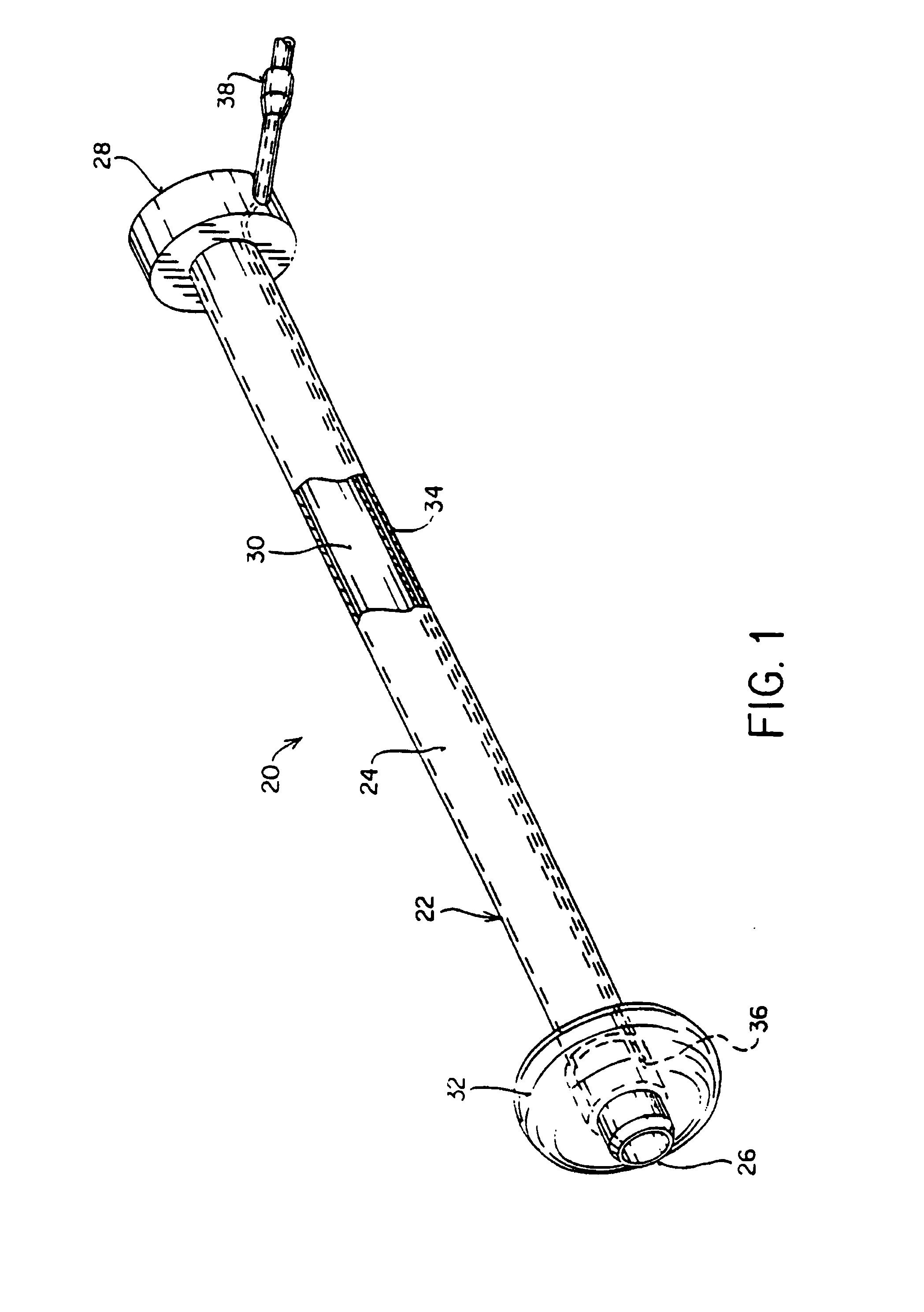
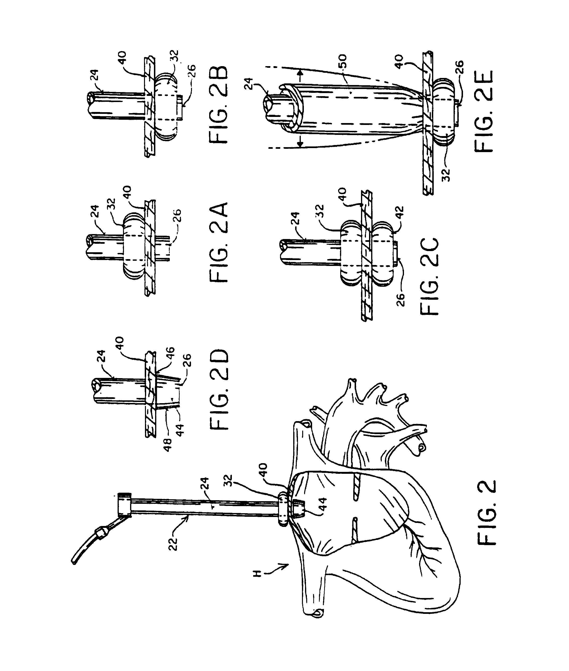
[0083]A first representative embodiment of an intracardiac access system according to the invention is illustrated in FIG. 1. The intracardiac access system 20 includes a tubular access device 22 comprising a rigid shaft 24 having a distal end 26, a proximal end 28, and an inner lumen 30 extending therebetween. Access device 22 includes a means near distal end 26 for hemostatically sealing a cardiac penetration through which shaft 24 is introduced, which may comprise a toroidal balloon 32. An inflation lumen 34 extends through shaft 24 and has an opening 36 in communication with the interior of balloon 32. An inflation fluid port 38 is mounted to shaft 24 at proximal end 28 in communication with inflation lumen 34 and is configured for connection to an inflation fluid delivery source such as a syringe or other balloon inflation device.

[0084]Access device 22 is configured to extend percutaneously through an intercostal space and through a muscular wall of the heart with distal end 26...


PUM


Abstract

Devices, systems, and methods are provided for accessing the interior of the heart and performing procedures therein while the heart is beating. In one embodiment, a tubular access device having an inner lumen is provided for positioning through a penetration in a muscular wall of the heart, the access device having a means for sealing within the penetration to inhibit leakage of blood through the penetration. The sealing means may comprise a balloon or flange on the access device, or a suture placed in the heart wall to gather the heart tissue against the access device. An obturator is removably positionable in the inner lumen of the access device, the obturator having a cutting means at its distal end for penetrating the muscular wall of the heart. The access device is preferably positioned through an intercostal space and through the muscular wall of the heart. Elongated instruments may be introduced through the tubular access device into an interior chamber of the heart to perform procedures such as septal defect repair and electrophysiological mapping and ablation. A method of septal defect repair includes positioning a tubular access device percutaneously through an intercostal space and through a penetration in a muscular wall of the heart, passing one or more instruments through an inner lumen of the tubular access device into an interior chamber of the heart, and using the instruments to close the septal defect. Devices and methods for closing the septal defect with either sutures or with patch-type devices are disclosed.

Description

CROSS-REFERENCE TO RELATED APPLICATIONS[0001]This application is a continuation of application Ser. No. 09 / 411,095, filed Oct. 4, 1999, now U.S. Pat. No. 6,401,720, which is a continuation of application Ser. No. 08 / 643,898, filed May 7, 1996, now issued as U.S. Pat. No. 6,079,414, which is a divisional of application Ser. No. 08 / 425,179, filed Apr. 20, 1995, now issued as U.S. Pat. No. 5,797,960, which is a continuation-in-part of application Ser. No. 08 / 163,241, filed Dec. 6, 1993, now issued as U.S. Pat. No. 5,571,215, which is a continuation-in-part of application Ser. No. 08 / 023,778, filed Feb. 22, 1993, now issued as U.S. Pat. No. 5,452,733. The complete disclosures of these applications are hereby incorporated herein by reference for all purposes.FIELD OF THE INVENTION[0002]The present invention relates generally to less-invasive surgery of the cardiovascular system. More specifically, the invention relates to thoracoscopic devices and techniques for performing surgical proce...

Claims


Application Information

Patent Timeline
Patent Type & Authority Patents(United States)
IPC IPC(8): A61B19/00A61B1/00A61B5/344A61B17/00A61B17/02A61B17/04A61B17/06A61B17/28A61B17/30A61B17/32A61B17/34A61B18/00A61B18/14A61F2/24A61F2/958A61M1/00
CPCA61B17/00234A61B17/0218A61B17/0469A61B17/06061A61B17/320016A61B17/3468A61B18/1482A61B18/1492A61B19/26A61F2/2427A61M25/1011A61M39/0247A61B17/0057Y10S623/904A61B17/0467A61B17/2909A61B17/3211A61B17/3417A61B18/1442A61B19/5225A61B2017/00053A61B2017/00243A61B2017/00247A61B2017/00349A61B2017/00575A61B2017/00579A61B2017/00592A61B2017/00597A61B2017/00606A61B2017/00619A61B2017/00623A61B2017/00867A61B2017/00907A61B2017/0243A61B2017/047A61B2017/0472A61B2017/0474A61B2017/0475A61B2017/2943A61B2017/2946A61B2017/306A61B2017/3405A61B2017/3486A61B2017/3492A61B2018/00214A61B2018/00232A61B2018/00261A61B2018/00291A61B2018/00363A61B2018/00392A61B2018/00577A61B2018/00898A61B2018/00982A61B2018/1465A61M2025/028A61M2039/0279A61M2039/0297A61M2205/3344A61M2205/3355A61M2205/366A61B90/50A61B90/37
Inventor STEVENS, JOHN H.REITZ, BRUCE A.ROTH, ALEX T.PETERS, WILLIAM S.GIFFORD, HANSON S.
Owner HEARTPORT
Why Eureka
Try Eureka