Image diagnosis supporting device

a supporting device and image technology, applied in the field of image diagnosis, can solve the problems of difficult use of the above-described image diagnosis supporting devi

Inactive Publication Date: 2007-11-20
FUJIFILM HEALTHCARE CORP
View PDF7 Cites 22 Cited by
  • Summary
  • Abstract
  • Description
  • Claims
  • Application Information

AI Technical Summary

Problems solved by technology

However, the above-described image diagnosis supporting device is difficult to use, because a lot of time-consuming work is required to adjust the parameters for specifying the sizes and shapes of various shadows.

Method used


Image

  • Image diagnosis supporting device
  • Image diagnosis supporting device
  • Image diagnosis supporting device

Examples


Embodiment Construction

[0095]Preferred embodiments of an image diagnosis supporting device according to this invention will be described with reference to the accompanying drawings.

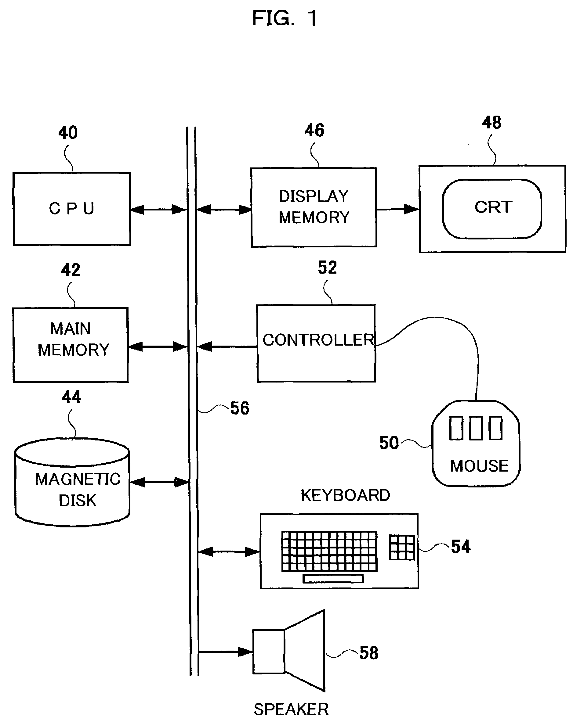
[0096]FIG. 1 is a block diagram showing the overall hardware construction of an image diagnosis supporting device to which this invention is applied. This image diagnosis supporting device displays extracted focus candidate shadows on the basis of a plurality of tomographic images (such as CT images) that are collected from a target area of a sample by means of, for example, an X-ray CT device. The image diagnosis supporting device selectively displays, in addition to focus candidate shadows, shadows of high certainty from among extracted focus candidate shadows or the like, or displays halfway images during this processing.

[0097]This image diagnosis supporting device is made up of a central processing unit (CPU) 40 which controls the operation of each constituent element, a main memory 42 in which a control program for the dev...


PUM


Abstract

An image diagnosis supporting device operates, through digitizing, to apply predetermined image processing to a medical image and to generate a multi-valued image having discrete multiple values. At least one decision making processing routine is then executed on the multi-valued image and/or the medical image; and, from among shadows in the processed image, a focus candidate shadow which is likely to indicate a diseased site is extracted. The extracted focus candidate shadow is then displayed in the medical image so that it is easily identifiable.

Description

TECHNICAL FIELD[0001]The present invention relates to an image diagnosis supporting device, which extracts a shadow that serves as a focus candidate (a possibly diseased portion) from a medical image by computerized image processing and displays the extracted shadow as is a focus candidate so that it can be identified.BACKGROUND OF THE INVENTION[0002]A computerized image diagnosis supporting device analyzes shadows in a medical image by using a computer, and displays a medical image containing a focus candidate shadow selected from the shadows, thereby presenting such medical image to a doctor who has been requested to make a diagnosis. The term “medical image” used herein covers photographic images photographed with medical image diagnosis devices, such as CT devices, MRI devices and ultrasonic diagnosis devices, as well as difference images between past images and current images and the like. As to the method of selecting a focus candidate, several examples associated with medical...

Claims


Application Information

Patent Timeline
Patent Type & Authority Patents(United States)
IPC IPC(8): G06K9/00A61B6/03A61B5/055G06T1/00G06T3/40G06T5/00G06T5/20G06T7/00G06T7/60
CPCA61B6/463A61B6/468G06T7/0012G06T7/0083A61B6/027G06T2207/10081G06T2207/30061G06T2207/30096G06T2200/24G06T7/12
Inventor GOTO, YOSHIHIRO
Owner FUJIFILM HEALTHCARE CORP
Why Eureka
Try Eureka