Method and apparatus for enhancing image acquired by radiographic system

a radiographic system and image technology, applied in image enhancement, image analysis, instruments, etc., can solve the problems of not meeting the requirements of relatively more enhancement, the resolving capability of contours in the dark region of the human eye is lower than that of contours, and the enhancement amplitude of weak edges is relatively small, so as to improve the resolving capability of contours in the dark region, eliminate image noise, and enhance the effect of image information

Active Publication Date: 2010-03-30
NUCTECH CO LTD +1
View PDF11 Cites 28 Cited by
  • Summary
  • Abstract
  • Description
  • Claims
  • Application Information

AI Technical Summary

Benefits of technology

[0006]In view of the above drawbacks in the prior arts, it is an object of the present invention to provide a method for enhancing image information in radiography, which may contrast-enhance edge details of various gradation regions in an image in a whole while eliminating image noises, and especially, increase an enhancement amplitude of weak edges in dark regions, in order to achieve a clear scanned image.
[0021]With the above technical solution, the present invention performs the enhancement process to images by using an adaptive non-linear contrast enhancement method. The adaptivity is characterized in that edge details of various gradation regions in an image are contrast-enhanced in a whole while eliminating image noises, and enhancement amplitude is increased at weak edges in dark regions, so as to make the scanned images clearer. As compared with the prior arts, the inventive method has a fast processing speed for image information enhancement and a simple algorithm, images clearly, eliminates noises in the images, and satisfies the requirements of relatively more enhancement to the contrast of the dark regions in the scanned images. The present invention is applicable to a processing system of image information enhancement with large data processing amount and high real-time requirements.

Problems solved by technology

Furthermore, human's eyes have lower resolving capability for contours in a dark region than that for contours in a bright region.
On the other hand, the enhancement to weak edges in the dark regions is relatively small, which can not satisfy the requirements of relatively more enhancement to the contrast of the dark regions in the scanned images.

Method used


Image

  • Method and apparatus for enhancing image acquired by radiographic system
  • Method and apparatus for enhancing image acquired by radiographic system
  • Method and apparatus for enhancing image acquired by radiographic system

Examples


Embodiment Construction

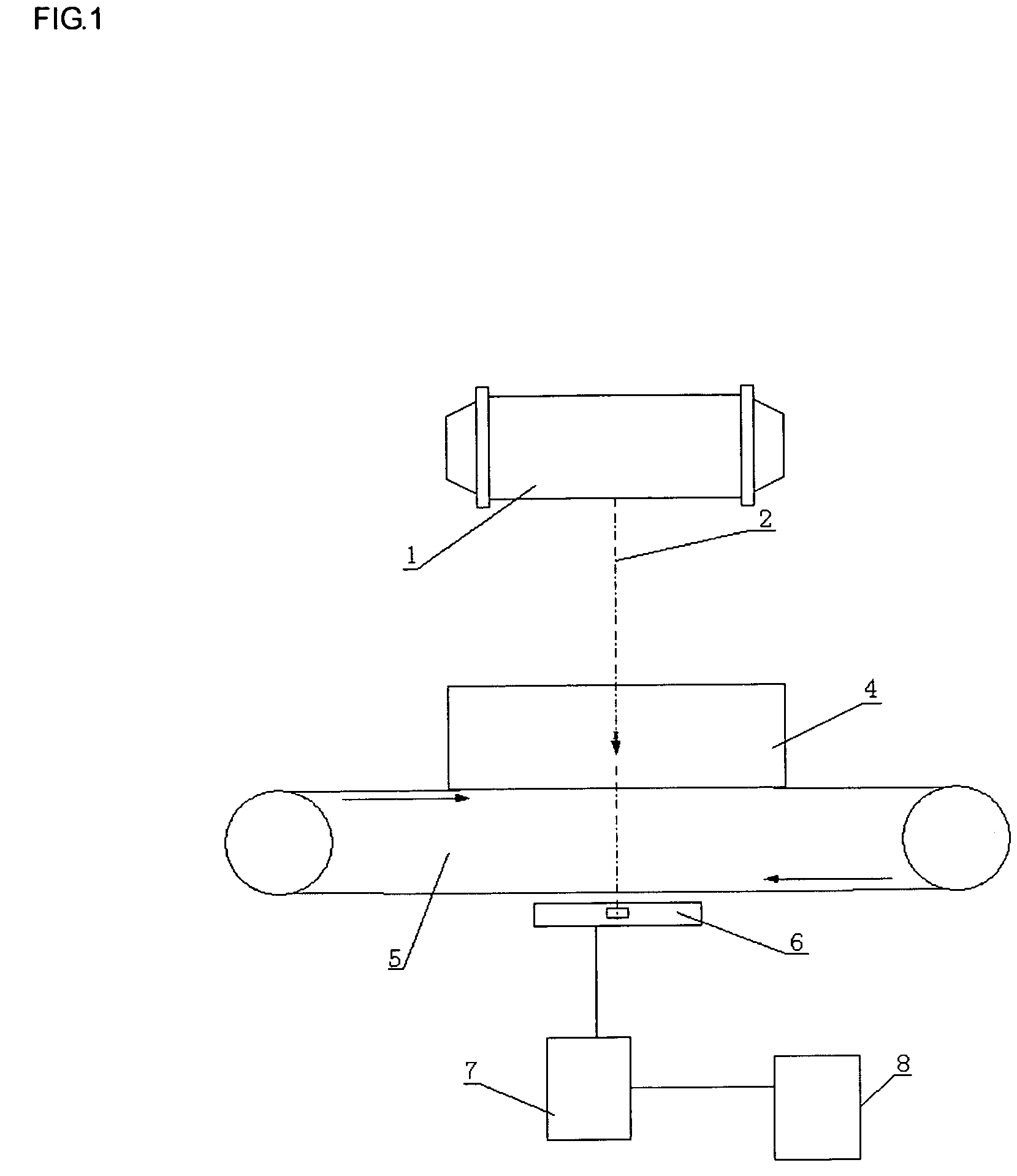
[0026]The inventive method is used for processing a signal obtained by a radiographic system. As shown in FIG. 1, the radiographic system includes a radiation source 1, a mechanical control device 5, a detector device 6, a data acquisition device 7 and a computer imaging device 8. The radiation source 1 emits X rays 2 which penetrate through an object to be detected 4 being carried by the mechanical control device 5, and then are received by the detector device 6 facing the X rays 2. The received X rays signal is transmitted from the detector device 6 to the data acquisition device 7, and after being converted into a digital signal, is transferred to the computer imaging device 8.

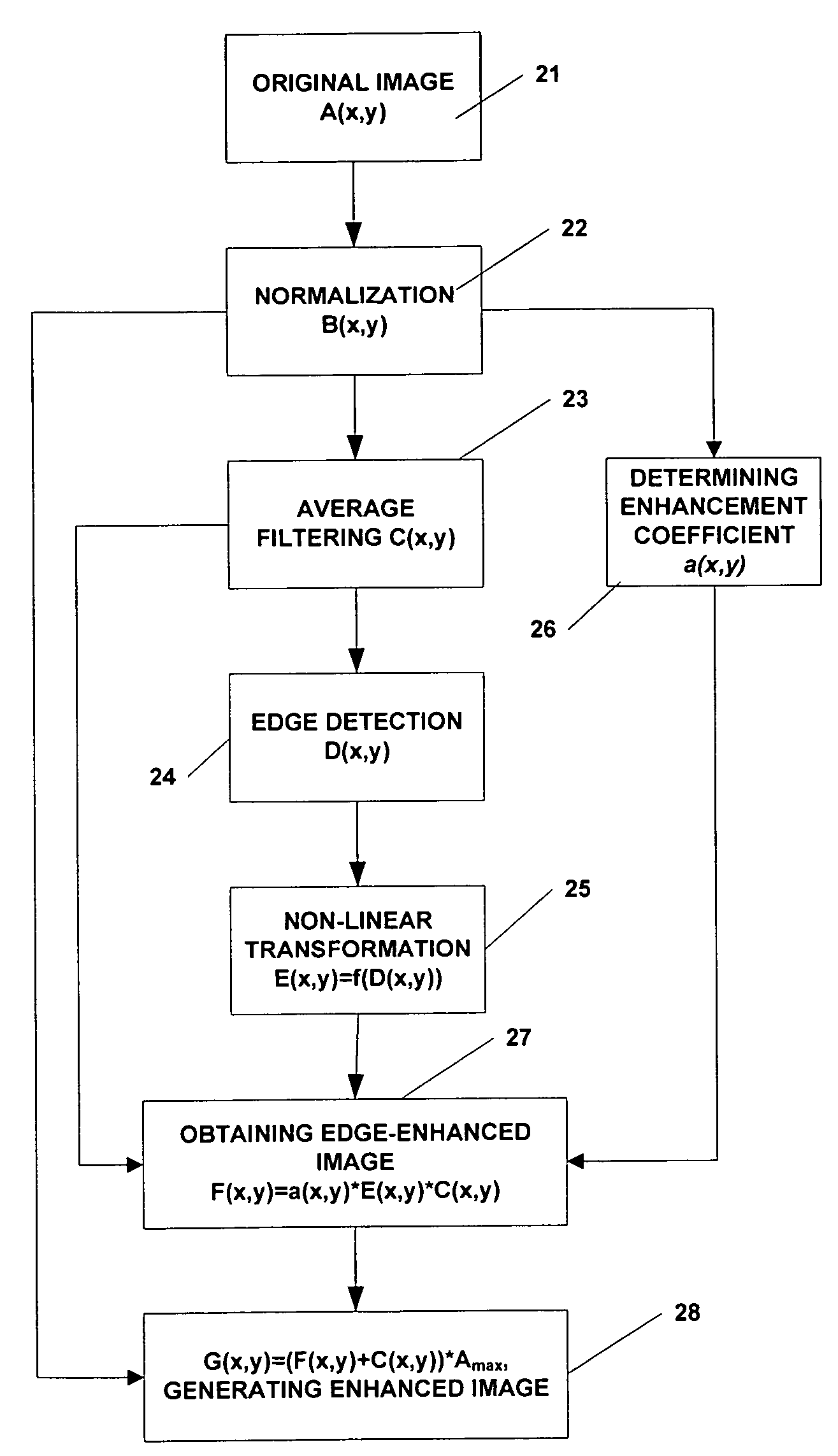
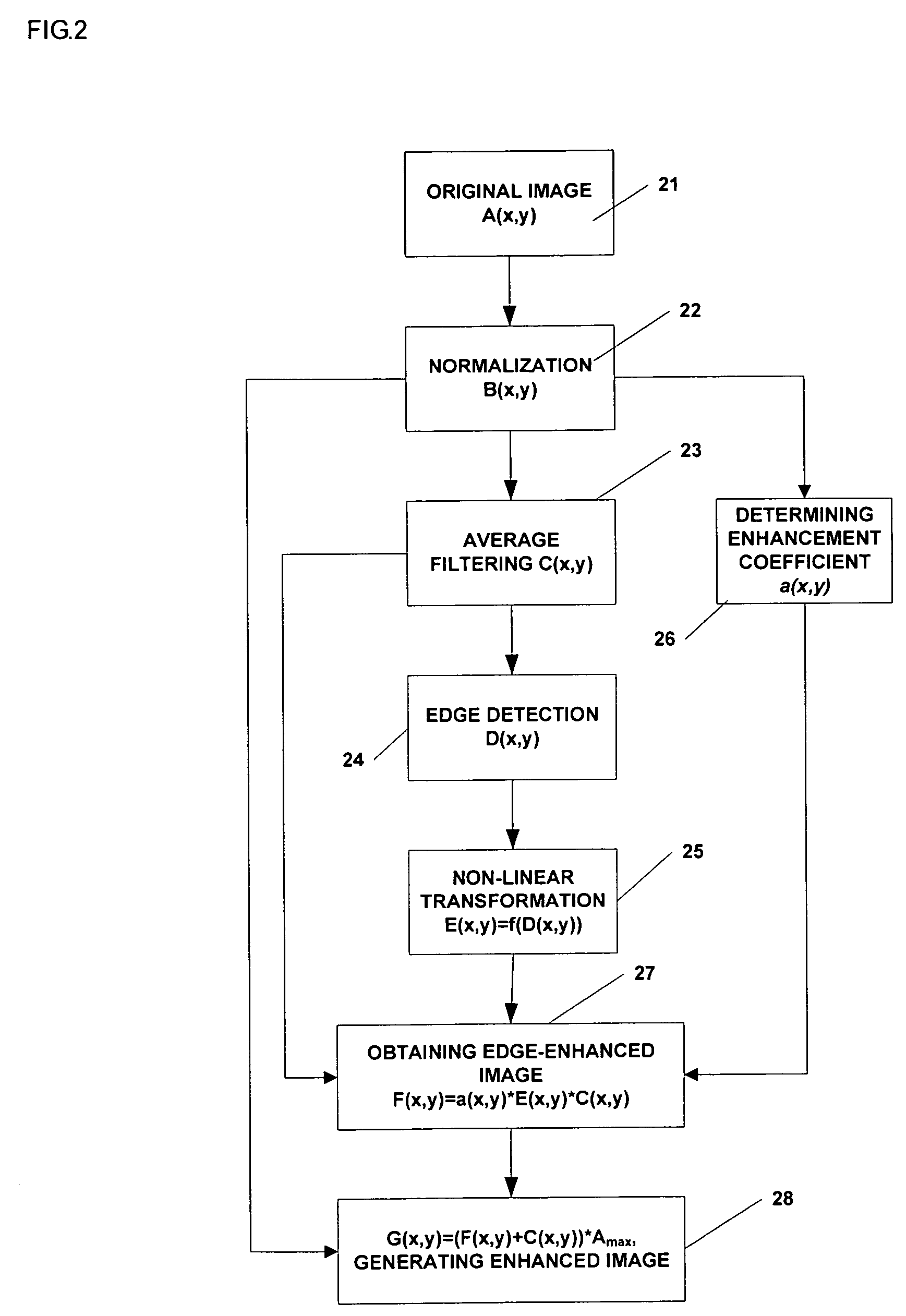
[0027]Referring to FIG. 2, the main steps of the inventive method are described as follows.

[0028]At block 21, a radiation image A(x,y) of the object to be detected is obtained through the radiographic system.

[0029]At block 22, a maximum value of pixel gradations Amax is found in the image A(x,y). Then, the ...


PUM


Abstract

A method of image information enhancement in radiography relates to image information processing techniques in radiography. The method comprising steps of: normalizing an acquired image A(x,y) to form a normalized image B(x,y); filtering the normalized image B(x,y) by a low-pass filter to obtain an filtered image C(x,y); calculating a relative standard deviation for each pixel in the image A(x,y), three times the relative standard deviation being an edge threshold for each pixel; thresholding a difference image obtained by subtracting the filtered image C(x,y) from the normalized image B(x,y) by using the edge threshold for each pixel to form a threshold-processed image D(x,y); enhancing a contrast of the threshold-processed image D(x,y) by using a non-linear function to form a contrast-enhanced image E(x,y); determining a enhancement coefficient a(x,y); obtaining a edge-enhanced image F(x,y) by multiplying the enhancement coefficient a(x,y), the contrast-enhanced image E(x,y) and the filtered image C(x,y); and generating a resulting image by multiplying a sum of the edge-enhanced image F(x,y) and the filtered image C(x,y) with the maximum value Amax As compared with the prior arts, the inventive method has a fast processing speed for image information enhancement and a simple algorithm, images clearly, eliminates noises in the images, and satisfies the requirements of relatively more enhancement to the contrast of the dark regions in the scanned images.

Description

BACKGROUND OF THE INVENTION[0001]1. Field of Invention[0002]The present invention relates to image information processing techniques in radiography, and more particularly to a method for enhancing image information in radiography.[0003]2. Description of Prior Art[0004]Radiographic technique is now widely used in industrial Nondestructive Detections (NDTs), medical diagnoses and science researches. The basic principle of radiography is that different absorptivities of different materials to X rays are used for forming different gradation values of pixels in a scanned image acquired by X rays penetrated an object. The energy of rays is exponentially attenuated when the thickness of the object to be scanned increases. Therefore, the difference among gradation values for border contours of a scanned image of a thicker object is much less than that of a thinner one, that is to say, the contours inside the object will be more blurred with increasing of the thickness of the object to be sc...

Claims


Application Information

Patent Timeline
Patent Type & Authority Patents(United States)
IPC IPC(8): G06K9/40
CPCG06T5/009G06T5/40G06T2207/10116G06T2207/30004
Inventor ZHANG, LICHEN, ZHIQIANGXING, YUXIANGLI, YUANJINGLIU, YINONGZHAO, ZIRANXIAO, YONGSHUN
Owner NUCTECH CO LTD
Why Eureka
Try Eureka