Two-photon endoscopic scanning assembly for inflammatory disease detection

a scanning assembly and endoscopic technology, applied in the field of optical instruments for imaging tissue using optical instruments, can solve the problems of limited benchtop systems, inability to achieve studies that would be impossible with a benchtop system, and inability to achieve studies that would be impossible with a benchtop system, and achieve the effect of large stroke length

Active Publication Date: 2014-08-19
RGT UNIV OF MICHIGAN
View PDF5 Cites 4 Cited by
  • Summary
  • Abstract
  • Description
  • Claims
  • Application Information

AI Technical Summary

Benefits of technology

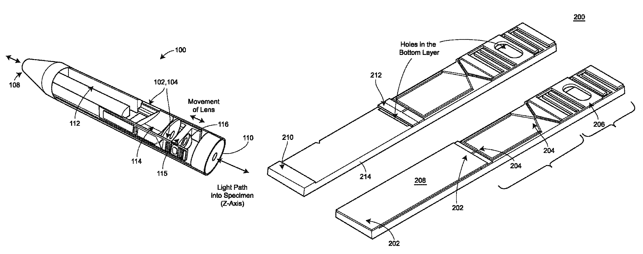
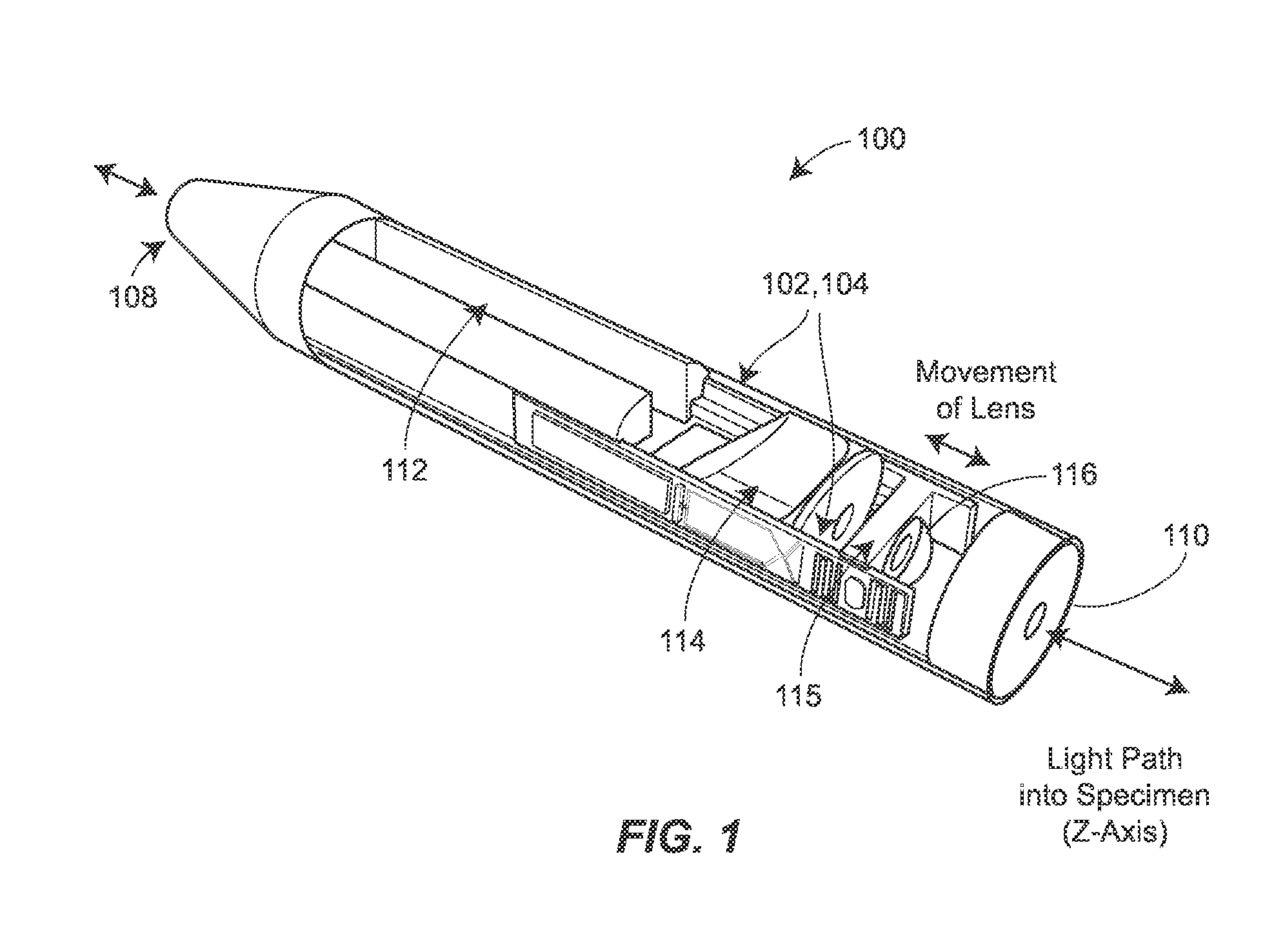
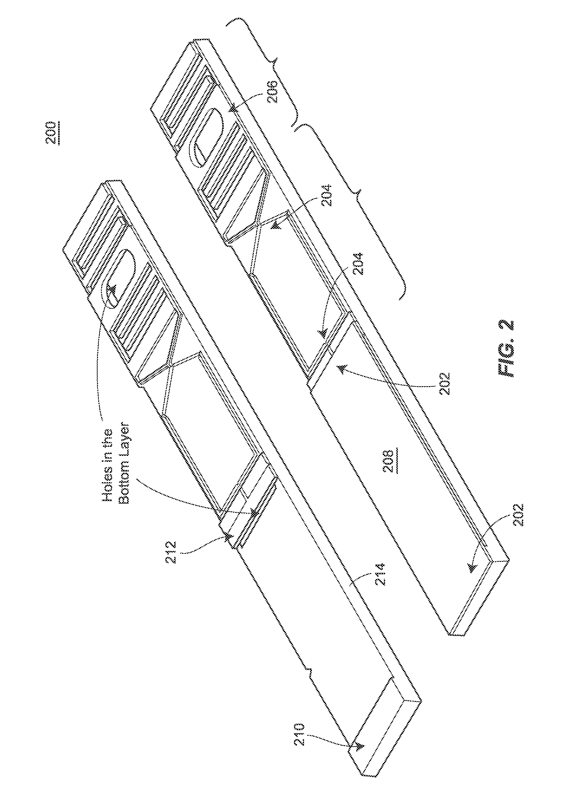
[0007]The actuators described herein may be used in any number of instruments and applications. For example, actuators may be paired with optical components to form an integrated device. These optical components may be integrated with one or more of the actuators using micromachined silicon flexures, to form a machined z-axis actuator. For example, actuators are described forming endoscopic instruments acting as two-photon microscopes, with an optical path occupying the center of the imaging system. Because the actuators are low-profile, they can be used in structures of typical endoscopic size, while still providing scanning depth ranges useful for microscopy. Actuators may be fit along the side of the optical path to minimize total cross-sectional area and volume of the scanning head used to generate images.

Problems solved by technology

By providing a platform for scanning image collection in a miniature device (e.g., in hand-held or endoscopic form) diagnosis, monitoring, and studies that would be impossible with a benchtop system may not be achieved.
Whereas, benchtop systems, by contrast, are limited to single time samples from biopsies, or specific locations on an animal that happen to “fit” into the system.
Further, other miniature microscopes, such as confocal microscopes, tend not to have as high resolution and are more prone to photo-bleaching and possibly disrupting the tissue, compared to short pulse, two-photon instruments as discussed herein.

Method used


Image

  • Two-photon endoscopic scanning assembly for inflammatory disease detection
  • Two-photon endoscopic scanning assembly for inflammatory disease detection
  • Two-photon endoscopic scanning assembly for inflammatory disease detection

Examples


example 1

[0072]In a first example implementation, bilateral nasal smears were performed on 30 human subjects with rhinitis symptoms, and imaged with a laboratory two-photon microscope with 162.5 mW excitation at 700 nm wavelength. Fluorescent images (see, e.g., FIG. 7) for emission between 500 and 600 nm were taken and compared to histology. A significantly greater mean fluorescence intensity was observed from eosinophils compared to epithelial cells, 13.8±4.3 versus 3.7±1.8 (p<0.01), respectively. A receiver operator curve (ROC) is shown in FIG. 8 presenting the sensitivity versus specificity at various threshold intensities for use of a two-photon excited fluorescence to distinguish eosinophils from epithelial cells, resulting in an area under the curve of 98%.

[0073]Using volume scanning (z-axis and xy-plane scanning), the present techniques were able to use the two-photon fluorescence technique to establish 3D volumetric imaging of eosinophils in esophageal mucosa, as shown in FIG. 9. In ...

example 2

[0076]In another study, patients aged 18-65 years who are undergoing routine endoscopy and have symptoms consistent with EoE, including dysphagia or food impaction were recruited for an endoscopic imaging analysis. Patients were excluded if they had a known bleeding disorder or an elevated International Normalized Ratio (>1.5) owing to anticoagulation. Patients with severe illness such as heart failure, difficulty breathing, or kidney failure were also excluded. A total of 23 patients were recruited into this study with ages ranging from 21 to 64 years old (mean 42±13), including 12 females and 11 males. The patient demographics, symptoms on presentation, therapy before the study, and cell count on multi-photon microscopy and histopathology are presented in Table 4.

[0077]

TABLE 4MultiphotonPathologyAgeGenderPresenting SymptomsTherapyAbsolute Eos#Max Eos#60FDysphagiaOmeprazole 40 mg bidDistal-13Distal-742MH / o impaction, stricture, dilationOmeprazole 20 mg prnNANA36MDysphagia, food imp...


PUM


Abstract

An endoscopic imaging device is described that achieves longitudinal axis (z-axis) scanning into a tissue or sample, using a piezoelectric microactuator. In some configurations, additional lateral (xy-plane) scanning is also achieved, to allow for the creation of full three-dimensional imaging, ex vivo or in vivo. The techniques may be used to image and diagnosis allergic rhinitis and eosinophilic esophagitis in tissue.

Description

STATEMENT REGARDING FEDERALLY SPONSORED RESEARCH OR DEVELOPMENT[0001]This invention was made with government support under CA136429 awarded by the National Institutes of Health. The government has certain rights in the invention.CROSS-REFERENCE TO RELATED APPLICATIONS[0002]The present application claims the benefit of U.S. Provisional Application No. 61 / 435,136, entitled “Two-Photon Endoscopic Scanning Assembly for Inflammatory Disease Detection,” filed on Jan. 21, 2011, which is hereby incorporated by reference herein in its entirety.FIELD OF THE DISCLOSURE[0003]The present disclosure relates generally to techniques for imaging tissue using an optical instrument and, more particularly, to techniques for allowing two-dimensional (2D) and three-dimensional (3D) scanning using an optical instrument.BACKGROUND[0004]The ability to perform endoscopic tissue imaging using a fiber-coupled two-photon laser has recently been demonstrated by several research groups. For many applications the ...

Claims


Application Information

Patent Timeline
Patent Type & Authority Patents(United States)
IPC IPC(8): F21V17/02A61B1/06
CPCA61B1/00172G01N21/6486A61B1/233G01N33/4833A61B1/0011A61B1/2733
Inventor OLDHAM, KENNWANG, THOMAS D.LIU, ZHONGYAOYE, JING YONG
Owner RGT UNIV OF MICHIGAN
Why Eureka
Try Eureka