Methods and compositions for the treatment or prevention of pathological cardiac remodeling and heart failure

A technology for cardiac remodeling and heart failure, applied in drug combinations, pharmaceutical formulations, organic active ingredients, etc., can solve the problems of unclear function of PDE1C, poorly documented expression and function, etc.

Inactive Publication Date: 2011-06-15
UNIVERSITY OF ROCHESTER
View PDF16 Cites 5 Cited by
  • Summary
  • Abstract
  • Description
  • Claims
  • Application Information

AI Technical Summary

Problems solved by technology

However, PDE1 expression and function in the heart are not well documented
PDE1C expression has been detected in human heart and cardiomyocytes (Vandeput et al., "Cyclic Nucleotide Phosphodiesterase PDE1C1 in Human Cardiac Myocytes," J Biol Chem. 282:32749-57 (2007)), however, the expression of PDE1C in human cardiomyocytes function is still unclear

Method used


Image

  • Methods and compositions for the treatment or prevention of pathological cardiac remodeling and heart failure
  • Methods and compositions for the treatment or prevention of pathological cardiac remodeling and heart failure
  • Methods and compositions for the treatment or prevention of pathological cardiac remodeling and heart failure

Examples


Embodiment 1

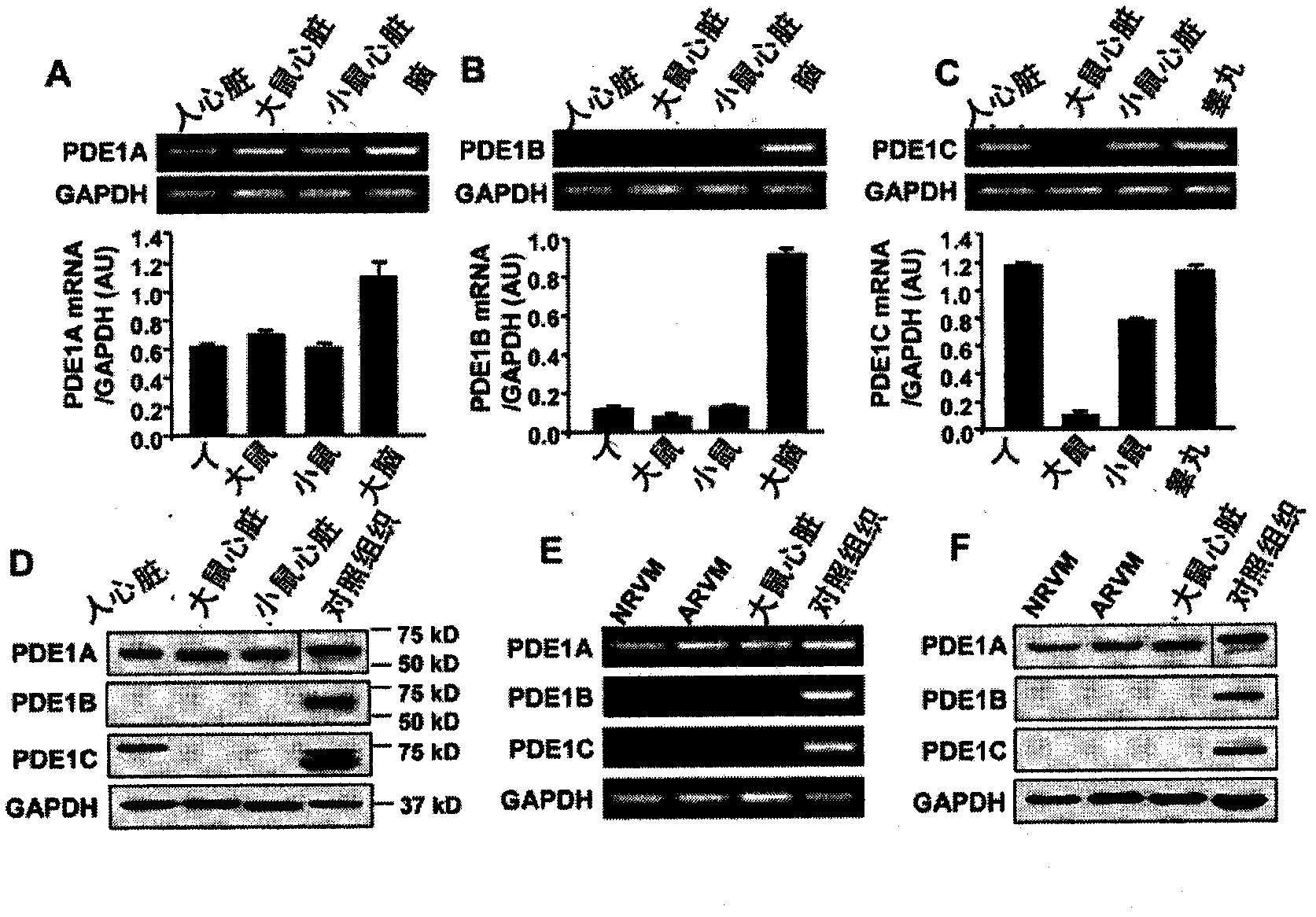
[0100] Example 1: Determining the expression of PDE1 isoforms in heart and cardiomyocytes

[0101] In human, rat, and mouse hearts, semi-quantitative RT-PCR analysis revealed that nearly identical levels of PDE1A were detected in human, rat, and mouse hearts, whereas PDE1C was predominantly detected in human and mouse hearts , PDE1B was weakly detected in all hearts ( figure 1 A-C). Western blot analysis showed that PDE1A protein levels were similar in hearts of different species, while PDE1B was not detected in hearts, consistent with mRNA expression ( figure 1 D). However, mouse hearts yielded much lower PDE1C protein expression compared to humans, inconsistent with mRNA expression levels ( figure 1 D). The low levels of mouse cardiac PDE1C protein are unlikely to be the result of antibody insensitivity, as the antibody recognizes mouse testis strongly ( figure 1 D). Furthermore, the levels of PDE1A mRNA and protein in both NRVM and ARVM were comparable to those in adult...

Embodiment 2

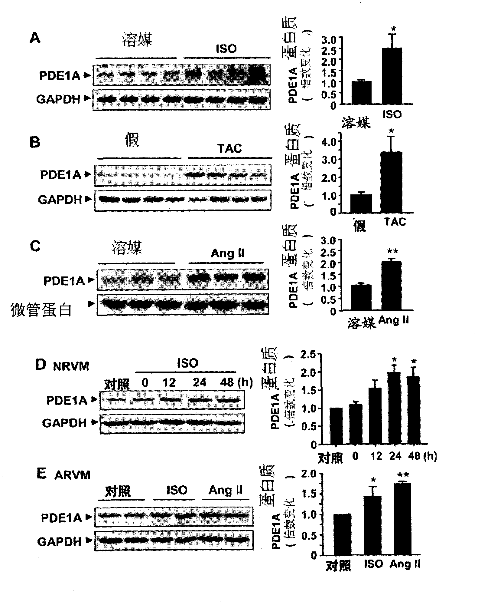
[0102] Example 2: PDE1A expression is upregulated in response to hypertrophic stimulation in isolated cardiomyocytes in vivo and in vitro

[0103] Western blot analysis showed that PDE1A protein levels were significantly upregulated in the hypertrophic hearts of animals, including the hearts of mice chronically infused (30 mg / kg / d, 7 days) with isoproterenol (ISO) ( figure 2 A) Hypertrophic heart of mice induced by chronic pressure overload after 4 weeks of transverse aortic constriction (TAC) ( figure 2 B) or rat hearts with long-term Ang II infusion (0.7 mg / kg / d, 7 days) via osmotic minipumps ( figure 2 C). These models are established rodent models of cardiac hypertrophy. In isolated NRVM, ISO treatment increased the relative level of PDE1A protein ( figure 2 D). Similarly, ISO or Ang II treatment of ARVM resulted in increased PDE1A protein levels ( figure 2 E). Collectively, these data indicate that PDE1A expression in cardiomyocytes can be upregulated by hypert...

Embodiment 3

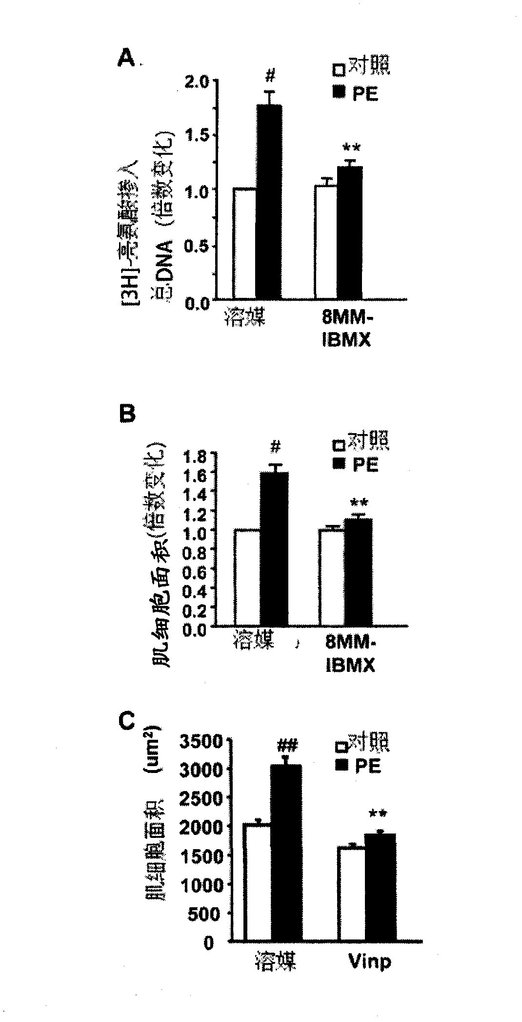
[0104] Example 3: Effect of PDE1 inhibition on hypertrophic growth of cardiomyocytes

[0105] pass 3 Protein synthesis by H-leucine incorporation ( image 3 A) or by muscle cell surface area ( image 3 B) Assessed, the PDE1 inhibitor 8-MM-IBMX (8-methoxymethyl-isobutylmethylxanthine) used at 20 μmol / L (a dose that selectively inhibits PDE1) significantly attenuated PE-induced neonatal Cardiomyocyte hypertrophy in rats. Measured by muscle cell surface area ( image 3 C), Vinpocetine (20 μM), known as a PDE1 inhibitor, also significantly reduced PE-induced myocyte hypertrophy. Neonatal rat cardiomyocytes were cultured in serum-free medium for 24 hours. Cells were pretreated with 20 μM of 8-MM-IBMX or vehicle DMSO, followed by either no treatment (control, ctrl) or treatment with PE for 48 hours. For the last 6 hours [ 3 H]-leucine-labeled pulse-chase. Cells were lysed, and the cell lysate was measured by scintillation counter 3 H-leucine incorporation. 3 Values ​​for H...


PUM


Abstract

The invention relates to methods of treating or preventing pathological cardiac remodeling and / or preventing heart failure. These methods include the administration of a PDEl inhibitor to a patient under conditions effective to treat or prevent pathological cardiac remodeling, and therefore heart failure that occurs as a result of such remodeling. Pharmaceutical compositions and delivery vehicles that can be used in the methods of the present invention are also disclosed herein.

Description

[0001] This application claims the benefit of US Provisional Patent Application Serial No. 61 / 050,308, filed May 5, 2008, the contents of which are hereby incorporated by reference in their entirety. field of invention [0002] The present invention relates to the use of PDE1 inhibitor for treating or preventing pathological myocardial remodeling and heart failure, and the pharmaceutical composition suitable for practicing these treatments or preventive treatments. Background of the invention [0003] Myocyte hypertrophy, caused by an increase in the size of individual cardiomyocytes, is key to physiological and pathological cardiac remodeling. Acquired hypertrophy during cardiac development or during exercise training is physiological hypertrophy and does not lead to decompensated heart failure. However, chronic mechanical and / or neurohumoral stress due to cardiovascular disease (eg, hypertension and myocardial infarction) induces excessive and persistent hypertrophy, which...

Claims


Application Information

Patent Timeline
Patent Type & Authority Applications(China)
IPC IPC(8): A61K31/404
CPCA61K31/4745A61P9/00A61P9/04
Inventor C·颜J-D·李
Owner UNIVERSITY OF ROCHESTER
Why Eureka
Try Eureka